| 图片: | |
|---|---|
| 名称: | |
| 描述: | |
- B1046Breast tubular leisons, MGA and differential diagnosis (cqz 2)
| 姓 名: | ××× | 性别: | F | 年龄: | 49 |
| 标本名称: | Breast excisional biopsy (乳腺切除活检) | ||||
| 简要病史: | |||||
| 肉眼检查: | |||||
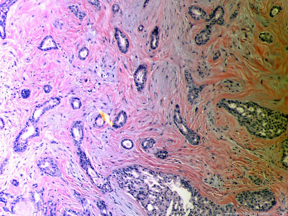
Microscopically it is a 0.8 cm lesion as photo.
Your diagnosis and differential diagnosis.
(镜下病变直径0.8cm,如图。请诊断和鉴别诊断)

名称:图1
描述:图1
标签:乳腺浸润性小管癌 硬化性腺病 微腺性腺病
-
本帖最后由 于 2010-05-16 23:38:00 编辑
相关帖子
- • 左乳肿物
- • 乳腺肿物一例
- • 腺病?癌?其他?(12楼常规,24楼免疫组化及会诊结果)
- • 左乳肿块,2X1.5cm,请会诊
- • 乳腺肿物
- • 求助:56岁女,左乳肿物,能排除小管癌吗?
- • 38岁乳腺(新加HE切片)
- • 乳腺包块。33岁
- • 乳腺肿块
- • 乳腺小管癌?
×参考诊断
1楼:SA,15楼:TC,22楼:非典型性MGA
-
本帖最后由 于 2008-12-24 20:14:00 编辑
| 以下是引用天山望月在2008-12-15 21:39:00的发言:
个人感觉: 仅从细胞图片看,有时不易诊断小管癌,需与导管内乳头状瘤或癌、DCIS、低级别的浸润性导管癌、淋巴瘤等鉴别。组织切片和免疫组化有助诊断。 不知对否?请赵老师点评!谢谢! |
You are right.
Like other low grade ca, TC is very difficult to make dx in FNA cytology, especially from the benign lesions. It is important you can call atypia and require core biopsy. Cell block may be helpful, but not IHC.


 重新阅读本主题,不好意思
 ,第一次没审清题,再次回答51楼的问题,并期待赵老师解答:


