| 图片: | |
|---|---|
| 名称: | |
| 描述: | |
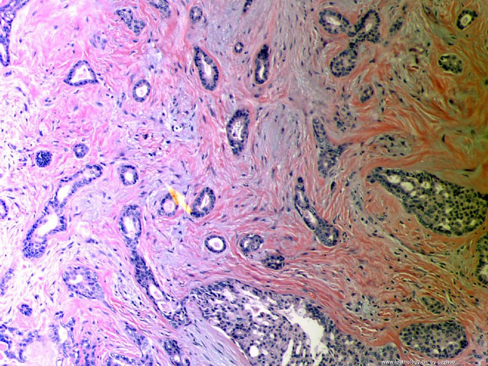
- B1046Breast tubular leisons, MGA and differential diagnosis (cqz 2)
| 姓 名: | ××× | 性别: | F | 年龄: | 49 |
| 标本名称: | Breast excisional biopsy (乳腺切除活检) | ||||
| 简要病史: | |||||
| 肉眼检查: | |||||
Microscopically it is a 0.8 cm lesion as photo.
Your diagnosis and differential diagnosis.
(镜下病变直径0.8cm,如图。请诊断和鉴别诊断)

名称:图1
描述:图1
-
本帖最后由 于 2010-05-16 23:38:00 编辑
相关帖子
- • 左乳肿物
- • 乳腺肿物一例
- • 腺病?癌?其他?(12楼常规,24楼免疫组化及会诊结果)
- • 左乳肿块,2X1.5cm,请会诊
- • 乳腺肿物
- • 求助:56岁女,左乳肿物,能排除小管癌吗?
- • 38岁乳腺(新加HE切片)
- • 乳腺包块。33岁
- • 乳腺肿块
- • 乳腺小管癌?
-
本帖最后由 于 2010-09-02 12:20:00 编辑
Am J Surg Pathol. 2009 Apr;33(4):496-504.
Molecular evidence for progression of microglandular adenosis (MGA) to invasive carcinoma.
Shin SJ, Simpson PT, Da Silva L, Jayanthan J, Reid L, Lakhani SR, Rosen PP.
Department of Pathology and Laboratory Medicine, New York-Presbyterian Hospital/Weill Cornell Medical Center, New York, NY 10065, USA. sjshin@med.cornell.edu
Abstract
Microglandular adenosis (MGA) is an uncommon, benign breast lesion that is characterized by a proliferation of small uniform, round glands lined by a single layer of epithelial cells around open lumina with haphazard infiltrative growth in fibrous and fatty breast tissue. Although MGA usually has an indolent course, there is morphologic evidence that MGA can be a precursor for the development of intraductal and invasive ductal carcinoma. To investigate the possibility of such a transition, we studied 17 cases of MGA or atypical MGA some of which had given rise to carcinoma in situ (CIS) and/or invasive ductal carcinoma using the reticulin stain, immunohistochemistry (S-100, p63, Ki-67, and p53), and a molecular approach involving microdissection and high-resolution comparative genomic hybridization and MYC chromogenic in situ hybridization. MGA and carcinomas arising from MGA were typically negative for p63 and positive for S-100 and Ki-67 and occasionally positive for p53. High-resolution comparative genomic hybridization identified recurrent gains and losses in MGA (2q+, 5q-, 8q+, and 14q-) and atypical MGA (1q+, 5q-, 8q+, 14q-, and 15q-). Some examples of MGA and carcinomas arising from MGA harbored few gross chromosomal abnormalities whereas others had considerable genetic instability with widespread aberrations affecting numerous chromosomal arms. Such widespread genetic changes, together with recurrent loss of 5q and gain of 8q were reminiscent of those reported specifically for basal-like, estrogen receptor-negative, and BRCA1-associated breast tumors. Concordant genetic alterations were identified between MGA, atypical MGA, and higher risk lesions (CIS and invasive ductal carcinoma) and in some cases there was an accumulation of genetic alterations as cases "progressed" from MGA to atypical MGA, CIS, and invasive ductal carcinoma. The molecular data suggests that MGA, atypical MGA, and carcinoma arising in MGA in a single case were clonally related. This result implicates MGA as a nonobligate precursor for the development of intraductal and invasive ductal carcinoma.


