


| 图片: | |
|---|---|
| 名称: | |
| 描述: | |
- B1046Breast tubular leisons, MGA and differential diagnosis (cqz 2)
| 姓 名: | ××× | 性别: | F | 年龄: | 49 |
| 标本名称: | Breast excisional biopsy (乳腺切除活检) | ||||
| 简要病史: | |||||
| 肉眼检查: | |||||
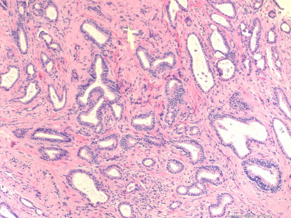
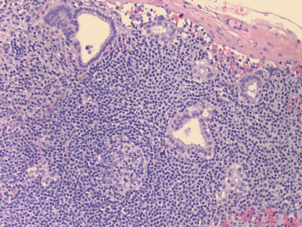
Microscopically it is a 0.8 cm lesion as photo.
Your diagnosis and differential diagnosis.
(镜下病变直径0.8cm,如图。请诊断和鉴别诊断)
图1")
名称:图1
描述:图1
标签:乳腺浸润性小管癌 硬化性腺病 微腺性腺病
-
本帖最后由 于 2010-05-16 23:38:00 编辑
相关帖子
- • 左乳肿物
- • 乳腺肿物一例
- • 腺病?癌?其他?(12楼常规,24楼免疫组化及会诊结果)
- • 左乳肿块,2X1.5cm,请会诊
- • 乳腺肿物
- • 求助:56岁女,左乳肿物,能排除小管癌吗?
- • 38岁乳腺(新加HE切片)
- • 乳腺包块。33岁
- • 乳腺肿块
- • 乳腺小管癌?
×参考诊断
1楼:SA,15楼:TC,22楼:非典型性MGA
-
本帖最后由 于 2008-10-12 22:12:00 编辑
Looks like tubular carcinoma, a well differentiated invasive ductal carcinoma. It requires >75%(?) tubular formation in neoplastic glands. Lower right hand portion of the photo shows possible low grade DCIS. The stroma probably represents desmoplastic response to those tumor glands. I talk with uncertain tone "probable, possible, looks like" because a single photomicrograph is a very limited source for a significant diagnosis, i cannnot be very sure.
abin译:
像小管癌,属于一种分化好的浸润性导管癌。它要求肿瘤性腺体中小管结构>75%(译注,这个标准有争议,有人严格要求100%。WHO认为小管结构达90%作为诊断标准可能较为妥当)。
右下可能为低级别DCIS。间质可能是促结缔组织反应。
-
本帖最后由 于 2008-10-12 22:56:00 编辑
 图1
图1 图2
图2 图3
图3 图4
图4
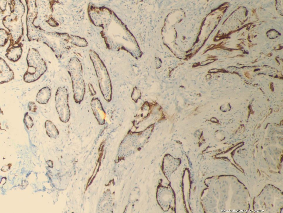
This is a typical tubular ca.
Fig 1 20x H&E
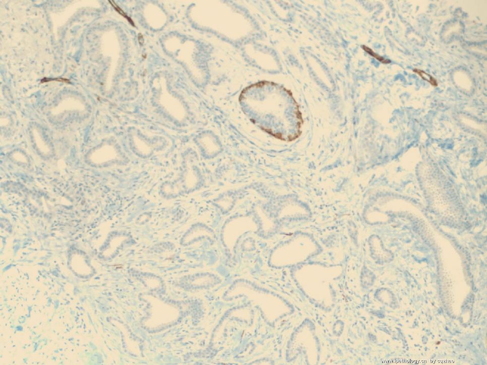
Fig 2.10x SMMHC stain
Fig 3. 10x P63 stain
Fig 4. 10x K67, very low proliferative index
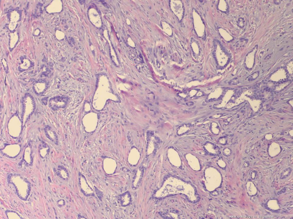
Tubular carcinoma (TC)is composed of small angular, oval, and tubular glands in the fibrotic background. Generally it is easy to make diagnosis. However it may be confused with sclerosing adenosis and microglandular hyperplasia (MGH).
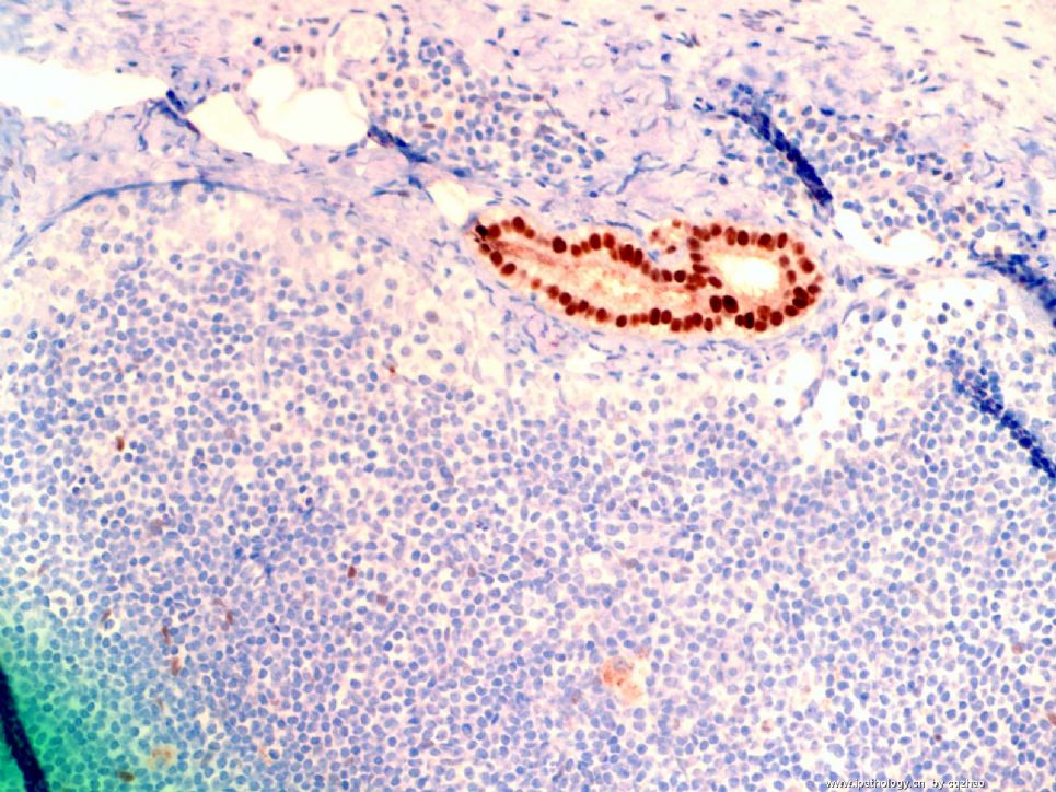
MGH glands also lack myoepithelial cells, but basement memberane stain (for example college IV) and S-100 stain are positive. These two stains are negative for TC. Microscopically, MGA is an infiltrative proliferation of small glands in fibrous or fatty stroma. MGA is composed of small uniform and regular glnads containing eosinophilic secretion.
It is not difficult to tell the difference between TC and sclerosing adenosis (SA). However, occasionally SA can be a mimic of TC. When the dx is unsure, stains of myoepithelial markers can be very useful like my first case.
Thank for reading
njwbhuang译:
第15楼提供的四张图,如下。这才是典型的小管癌。
小管癌是由一些小的有角的、卵圆形和小管状腺体组成,包埋于纤维性间质背景中。一般情况下较易诊断,然而它可与硬化性腺病和微腺体增生(MGH)混淆。
MGH中腺体也无肌上皮细胞,但基底膜染色(Ⅳ型胶原)和S-100蛋白染色阳性,而小管癌中则染色阴性。镜下,微腺性腺病(MGA)是小的腺体浸润于纤维或脂肪间质内,腺体较小,大小较一致,腺腔内含有嗜酸性分泌物。
小管癌与硬化性腺病(SA)的鉴别并不困难。然而,偶尔SA可能误诊为小管癌。当诊断不确定时,正如本例一样,肌上皮标记物有助于诊断。
谢谢阅读。
-
stevenshen 离线
- 帖子:343
- 粉蓝豆:2
- 经验:343
- 注册时间:2008-06-03
- 加关注 | 发消息
-
本帖最后由 于 2008-10-12 22:41:00 编辑
Thanks very much for this very challenging case and great discussion. I have to say that I would also make the diagnosis of invasive tubular carcinoma as well by looking at the first picture. It appears to have all the features that we use for a tubular carcinoma. I am interested to know why Dr. Zhao even consider to do the immunostain in the first place. Can you please share with us your diagnostic process and experience. What morphologic features that prompt you to order an IHC? If I don't do the stain, I would make a wrong diagnosis. It is true that tubular carcinoma is one of the many common pitfalls in breast pathology. Thanks again.
abin译:
非常感谢提供如此具有挑战性的病例,并有极好的讨论。我一开始也把第一张图诊断为浸润性小管癌了。它似乎具有我们用于诊断小管癌的所有特征。我很想知道,Dr. Zhao为什么第一步会想到做免疫组化。能否请教诊断过程和经验?什么形态特征促使您作IHC?我如果不作IHC,可能会误诊。确实,小管癌是乳腺病理诊断中的许多常见陷阱之一。再次感谢。
-
本帖最后由 于 2008-10-12 22:54:00 编辑
Lower Power: Generally, typical tubular carcinomas show angular and irregular glands with open lumens in desmoplastic stroma, such as my previous two cases. Sclerosing adenosis can also exhibit varying numbers of round, oval, or angular glands with open lumen. However the findings of open lumen often are not obvious.
High power: The nuclei of TC are often round and uniform, but not for SA.
Please remember that breast bx, skin malanoma, and pap test are the top three for lawsuit. Unfortunately I am a gyn/breast apthologist and need to deal with the two areas of the three every day. Anyway, You have to think over before you release any malignant case.
Hope it can help. Thnaks.
abin译:小管癌和硬化性腺病的鉴别要点
低倍:成角和不规则的腺体,管腔开放,促结缔组织增生性间质,正如我已经上传的两个病例。硬化性腺病(SA)也表现为不同数量的圆形或卵圆形或成角腺体,管腔开放。然而,开放的管腔通常不明显。
高倍:小管癌的核通常圆而一致,而SA不是这样。
请记住,乳腺活检,皮肤恶黑,和宫颈细胞学引起的诉讼最多。不幸得很,我从事妇科/乳腺病理,每天都需要处理三者中的两个。总之,发出恶性诊断之前,一定要再三推敲。
希望有帮助。谢谢。
-
本帖最后由 于 2008-10-12 22:22:00 编辑
To the Second floor:
There are no consensus concerning the proportion of tubular structures to establish the dx of TC. Some suggest the different cut-off points 75%, 90%, 100%. New WHO mentioned that for pragmatic reason, a 90% purity requirement offers a practical solution. For my daily practie, I use 100% as cut off point. Otherwise I call invasive ductal carcinoma with (focal) tubular feature. Few percentage of high grade carcinoma may cause poor prognosis.
In the right side, few ducts show UDH and no ADH present.
abin译:
关于诊断所需的小管结构的比例,尚未一致,有三种观点75%,90%和100%。新WHO提出90%作为实用标准。
我在日常工作中采用100%的诊断标准。否则我称为浸润性导管癌伴(局灶)小管癌特征。很少比例的高级别癌也可能预后差。
切片右侧一些导管为UDH,没有ADH。
-
本帖最后由 于 2008-10-12 22:34:00 编辑
 图1
图1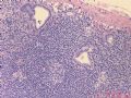 图2
图2 图3
图3
Just released another tubular carcinoma and share with people who love breast pathology.
Slide Key:
H&E 10x
Lymph node 20x
Lymph node 20x ER stain
This is a very low grade tumor of 0.7 cm with Nottingham grade 1. However axillary SNB shows 1 mm micrometastases.
Definition:
Isolated tumor cell: single cells or small clusters of cell not greater than 0.2 mm. (孤立的肿瘤细胞:单个细胞或小簇,不超过0.2mm)
Micrometastases: >0.2 mm to <2 mm. pN1mi (微小转移:>0.2mm 但<2 mm)
Thanks


