| 图片: | |
|---|---|
| 名称: | |
| 描述: | |
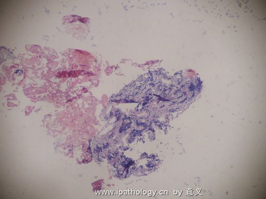
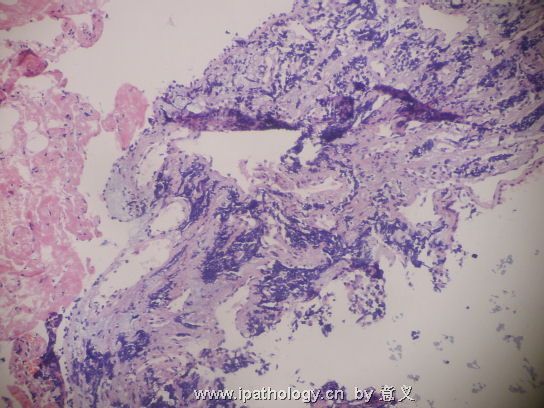
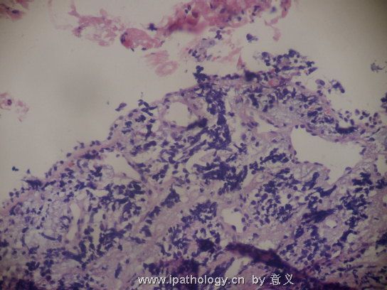
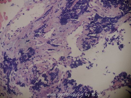
- B779请会诊,肺小细胞癌?
| 姓 名: | ××× | 性别: | 男 | 年龄: | 61 |
| 标本名称: | 右下支气管粘膜。 | ||||
| 简要病史: | 咳嗽月余,CT示肺癌,纤支镜见右下支气管外压肿物, | ||||
| 肉眼检查: | 镜下细胞挤压较重,能直接发癌吗? | ||||
名称:图1
描述:图1
名称:图2
描述:图2
名称:图3
描述:图3
名称:图4
描述:图4
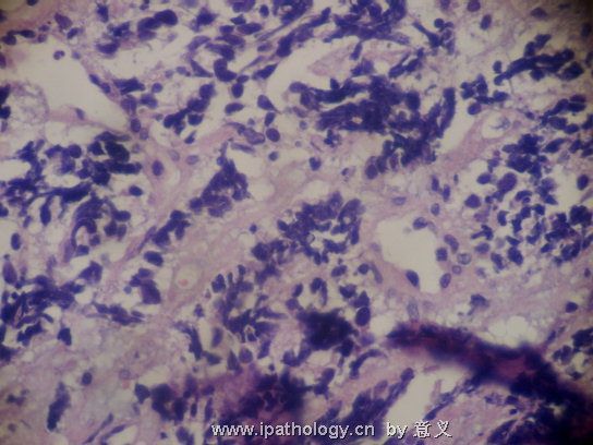
名称:图5
描述:图5
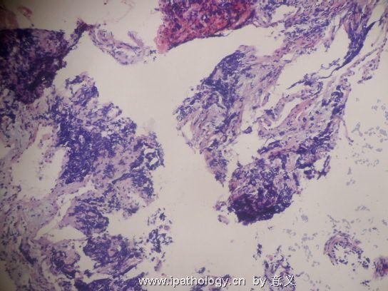
名称:图6
描述:图6
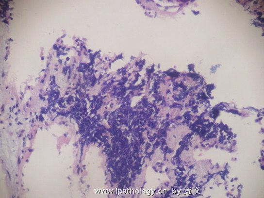
名称:图7
描述:图7
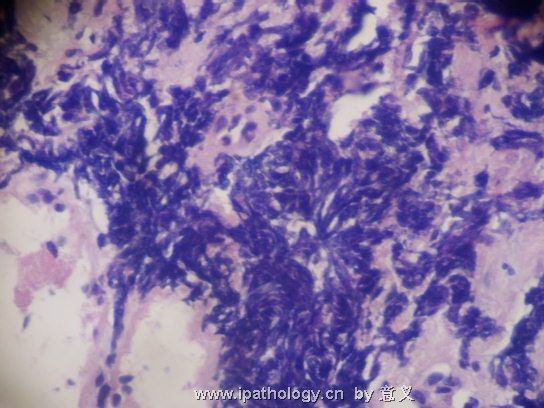
名称:图8
描述:图8

- 干到老学到老
相关帖子
-
本帖最后由 于 2006-11-20 16:38:00 编辑
| 以下是引用一笑 在2006-11-4 21:19:00的发言: 谢谢月新老师的翻译,我的英语底子太差,英文贴子我不大看得懂;象这个病例,乍看似乎有问题的,再看又不清楚,这种情况我们一般是要推给临床的,担子病理科不挑;如果要做免疫组化鉴别,等于已经承认是恶性肿瘤了,假若做一大堆组化,结果再不理想就很难和病人沟通,临床也会把责任推过来,被投诉的几率就非常高;我们一旦被投诉一次,这一年的安全奖就扣完了...... |
小心点, 没有100%的把捂, 不要发.
一个很有用的东西是KI67. 因为细胞积压太厉害, 形态上不好看.
小细胞癌是KI67>50%, 而类癌症<20%) Ki-67 . ROSAI 前几年写过一篇文章.
Am J Surg Pathol. 2005 Feb;29(2):179-87.
Typical and atypical pulmonary carcinoid tumor overdiagnosed as small-cell carcinoma on biopsy specimens: a major pitfall in the management of lung cancer patients.
Division of Pathology and Laboratory Medicine, European Institute of Oncology, University of Milan School of Medicine, Milan, Italy. giuseppe.pelosi@ieo.it
Seven patients with typical or atypical pulmonary carcinoid tumors overdiagnosed as small-cell carcinoma on bronchoscopic biopsies are described. Bronchial biopsies from 9 consecutive small-cell lung carcinoma patients were used as control group for histologic and immunohistochemical studies (cytokeratins, chromogranin A, synaptophysin, Ki-67 [MIB-1], and TTF-1). The carcinoid tumors presented as either central or peripheral lesions composed of tumor cells with granular, sometimes coarse chromatin pattern, high levels of chromogranin A/synaptophysin immunoreactivity, and low (<20%) Ki-67 (MIB-1) labeling index. The tumor stroma contained thin-walled blood vessels. Small-cell carcinomas always showed central tumor location, finely dispersed nuclear chromatin, lower levels of chromogranin A/synaptophysin, and high (>50%) Ki-67 (MIB-1) labeling index. The stroma contained thick-walled blood vessels with glomeruloid configuration. Judging from this study, overdiagnosis of carcinoid tumor as small-cell carcinoma in small crushed bronchial biopsies remains a significant potential problem in a worldwide sample of hospital settings. Careful evaluation of hematoxylin and eosin sections remains the most important tool for the differential diagnosis, with evaluation of tumor cell proliferation by Ki-67 (MIB-1) labeling index emerging from our review as the most useful ancillary technique for the distinction.
-
huaxiaxzmc 离线
- 帖子:229
- 粉蓝豆:24
- 经验:568
- 注册时间:2006-11-06
- 加关注 | 发消息
-
曹大夫认为:典型和非典型类癌很易过诊为小细胞癌,在活检标本中:在肺癌处理病人中的一个主要的陷井.
文献中共报道7例这样过诊的病例.类癌可表现为中央型或周围性病变.肿瘤细胞呈腺样,染色质颗粒状或粗糙.高水平的 chromogranin A/synaptophysin immunoreactivity,和低水平的 (<20%) Ki-67 (MIB-1) 标记指数.肿瘤间质包含薄壁血管.小细胞癌总是显示中央型定位,细而均匀分布的染色质.低度水平的chromogranin A/synaptophysin,和高水平的(>50%) Ki-67 (MIB-1) 增殖指数.间质含厚壁血管并呈球样构型.在小的挤压纤支镜活检标本中类癌过诊为小细胞癌目前仍是全球性问题.. 仔细观光HE切片及估计Ki-67 (MIB-1) 标记指数是鉴别的最有效的工具.(文献试译其主要观点).

朱正龙


