| 图片: | |
|---|---|
| 名称: | |
| 描述: | |
- B1046Breast tubular leisons, MGA and differential diagnosis (cqz 2)
| 姓 名: | ××× | 性别: | F | 年龄: | 49 |
| 标本名称: | Breast excisional biopsy (乳腺切除活检) | ||||
| 简要病史: | |||||
| 肉眼检查: | |||||
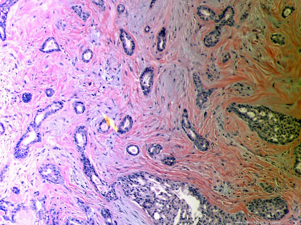
Microscopically it is a 0.8 cm lesion as photo.
Your diagnosis and differential diagnosis.
(镜下病变直径0.8cm,如图。请诊断和鉴别诊断)

名称:图1
描述:图1
-
本帖最后由 于 2010-05-16 23:38:00 编辑
相关帖子
- • 左乳肿物
- • 乳腺肿物一例
- • 腺病?癌?其他?(12楼常规,24楼免疫组化及会诊结果)
- • 左乳肿块,2X1.5cm,请会诊
- • 乳腺肿物
- • 求助:56岁女,左乳肿物,能排除小管癌吗?
- • 38岁乳腺(新加HE切片)
- • 乳腺包块。33岁
- • 乳腺肿块
- • 乳腺小管癌?
回22楼和26楼:
需要在微腺性腺病和浸润癌之间鉴别。腺腔小,较一致,腺体周围似有肌上皮混杂,支持微腺性腺病。但部分腺上皮异型性明显,关键是脂肪组织中有浸润。不能除外癌。必须做P63,CK5/6和SMA等肌上皮标记。
期待Dr. Zhao的讲解。。。。。。

- If you have great talents, industry will improve them; if you have but moderate abilities, industry will supply their deficiency. 如果你很有天赋,勤勉会使其更加完美;如果你能力一般,勤勉会补足其缺陷。
-
stevenshen 离线
- 帖子:343
- 粉蓝豆:2
- 经验:343
- 注册时间:2008-06-03
- 加关注 | 发消息
-
本帖最后由 于 2008-10-12 23:18:00 编辑
Florid small glandular proliferation with infiltration of stroma and fat = microglandular adenosis pattern; marked cytologic atypia including prominent nucleoli; stroma alteration more than that of benign microglandular adenosis. Morphologic diagnoses infiltrating ductal carcinoma with microglandular adenosis pattern or arising from microglandular adenosis. IHC stain with myoepithelial markers and collagen IV and S100 (as described by Dr. Zhao) will be help to confirm the diagnosis. Never seen such a case. Look forward to hearing about the final diagnosis. Thanks.
abin译:小腺体旺炽性增生,浸润间质和脂肪,这是微腺腺病的生长方式;有明显的细胞学非典型性,包括明显核仁;间质的改变也超出了良性微腺腺病的程度。
形态学诊断:呈微腺腺病结构的浸润性导管癌,或发生于微腺腺病的浸润性导管癌。
作肌上皮标记和S-100免疫组化以及胶原IV染色有助于确诊。从未见过这样的病例。期待最后诊断。
谢谢。


