| 图片: | |
|---|---|
| 名称: | |
| 描述: | |
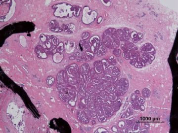
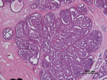
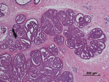
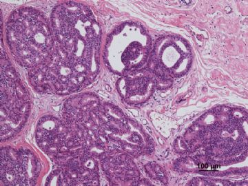
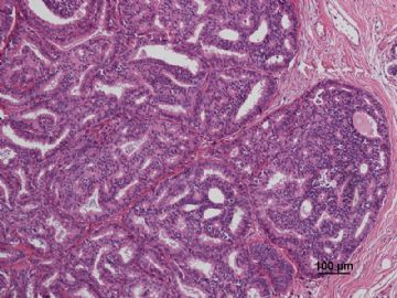
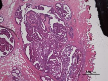
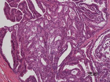
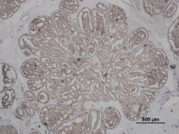
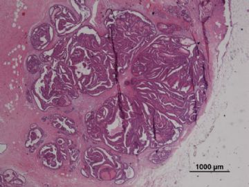
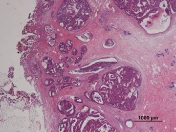
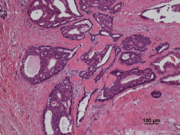
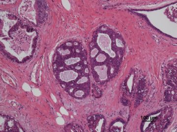
- B3087有难度的乳头状肿瘤病例(非典型性周围型导管内乳头状瘤还是周围型导管内乳头状瘤伴DCIS?)新加病灶测..
| 姓 名: | ××× | 性别: | F | 年龄: | 37 |
| 标本名称: | |||||
| 简要病史: | |||||
| 肉眼检查: | |||||
病历摘要:
“发现左乳肿物1年余,右乳肿物1周”入院。
2010年12月6日双乳钼靶片示:双乳呈混合型Ⅳb(纤维囊性增生为主),左乳外上区多发肿块不除外恶变可能,建议手术,右乳外上区良性增生结节,BI-RADS:III,左乳BI-RADS:V;
查乳腺彩超示:符合双侧乳腺增生声像,左乳头外上象限低回声团块,考虑为实性占位,不除外恶变可能,建议病理检查,右乳低回声肿块,考虑纤维腺瘤可能。
专科检查:双乳外形欠对称,左乳12-2点位距乳头2cm可触及一肿物,大小约4.0×5.0cm,质硬,边界欠清,表面欠光滑,活动度欠佳,与皮肤、胸壁无粘连。右乳11点位距乳头2cm可触及一肿物,大小约1.5×1.5cm,质韧,边界尚清,表面尚光滑,活动度一般,与皮肤、胸壁无粘连。
临床诊断:
1.乳房肿物(左乳癌?)2.乳房肿物(右乳增生结节?)3.乳腺增生(双侧)
大体病理检查:
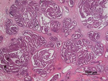
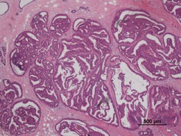
送检(左侧)乳腺肿物切除标本:6.0cm×4.0cm×1.0cm,切面见大小为1.5cm×1.3cm灰红结节。
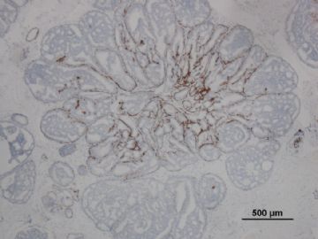
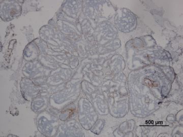
其中一张切片,免疫组化顺序为:ER,CK5/6,CD10
-
本帖最后由 于 2011-01-07 21:28:00 编辑
相关帖子
- • 乳腺包块
- • 左乳癌标本乳头一个导管内的病变
- • 乳腺两个相邻导管内的病变
- • 乳腺肿物
- • 乳腺肿物,请各位老师帮忙会诊
- • 女 46岁发现左乳腺肿块一月余
- • 乳腺包块。33岁
- • 左乳肿块,协助诊断
- • 乳腺肿物
- • 乳腺肿物
-
本帖最后由 于 2011-03-20 23:39:00 编辑
Papillomas can display focal proliferations of a mildly
atypical, monotonous cell population identical to lowgrade
DIN (DIN 1/ADH/DCIS, grade 1; Figures 5, A
through F, and 6, A through C). When DIN 1 occupies
less than a third of the papillary lesion, the term atypical
papilloma has been used. If at least a third, but less than
90% of the lesion displays such changes, the designation
of carcinoma arising in a papilloma has been used.
When completely excised, these 2 groups do not seem to differ in clinical behavior, as observed in a retrospective study that used this quantitative approach. It is noteworthy that
although 90% was used as the cut-off point, none of the
lesions had atypical areas that occupied more than 65%
to 70% of the papillary lesion. Currently, to avoid the term
carcinoma, we designate all such lesions as papillomas with
DIN 1/atypical papilloma (Table 1).
-Ueng, S.H., T. Mezzetti, and F.A. Tavassoli, Papillary neoplasms of the breast: a review. Arch Pathol Lab Med, 2009. 133(6): p. 893-907.

