共4页/67条首页上一页1234下一页尾页
回复:67 阅读:7909
头痛脑脊液检查

cqzhao 离线

帖子:5518
粉蓝豆:1071
经验:5755
注册时间:2008-09-29
加关注  |  发消息
楼主 发表于 2010-02-13 10:34|举报|关注(0)
浏览排序[ 顺序 逆序 楼主 支持 精彩 ]  快捷回复

Our fellow showed a case last week. I think it is interesting. Send here as a chinese new year gift for some ones interested.

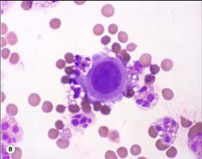
about 50 y women with headach, CSF cytology exam. paitent has no previous malignant history.

  • 头痛脑脊液检查图1
    图1
  • 头痛脑脊液检查图2
    图2
  • 头痛脑脊液检查图3
    图3
  • 头痛脑脊液检查图4
    图4
标签:
本帖最后由 于 2010-02-13 10:36:00 编辑
0
×参考诊断
最后诊断:恶性黑色素瘤,结合临床,可能原发于脑膜。

海上明月 离线

帖子:9476
粉蓝豆:1172
经验:10007
注册时间:2009-08-29
加关注  |  发消息
41 楼    发表于2010-03-28 19:35:00举报|引用
返回顶部 | 快捷回复

 细胞形态不规则,高核/浆比,大核仁(1-2个/每个细胞),胞浆内偶见颗粒状物(色素颗粒?)。

结合免疫细胞化学标记和讨论中Dr.Zhao的有关病史陈述,考虑为脑膜来源黑色素细胞病变:(1)黑色素细胞增生症恶性变?(2)黑色素细胞瘤(melanocytoma)进展?(3)恶性黑色素瘤?(4)中间型或混合性黑色素细胞肿瘤?

0
回复
signature
王军臣

海上明月 离线

帖子:9476
粉蓝豆:1172
经验:10007
注册时间:2009-08-29
加关注  |  发消息
42 楼    发表于2010-03-28 19:42:00举报|引用
返回顶部 | 快捷回复
本帖最后由 于 2010-03-28 20:44:00 编辑  综上述,不管这例黑色素病变是否有混合,脑脊液中能查出恶性特征的细胞,考虑为恶性黑色素瘤,倾向脑膜来源
0
回复
signature
王军臣

海上明月 离线

帖子:9476
粉蓝豆:1172
经验:10007
注册时间:2009-08-29
加关注  |  发消息
43 楼    发表于2010-03-28 20:42:00举报|引用
返回顶部 | 快捷回复
以下是引用cqzhao在2010-2-23 3:55:00的发言:


If it is multiple choice question, which one will you pick?


The best interpretation of the CSF findings is:

A.  Negative, reactive monocytes
B.  Atypical cells present
C. Suspicious for malignancy
D. Positive for malignant cells, epithelial vs. non-epithelial malignancy
E. Positive for malignant cells, adenocarcinoma


多选题,选其一。

本例脑脊液检查所见的最佳表述是:

A.阴性,考虑为反应性单(个)核细胞

B.可见非典型性细胞

C.疑恶性肿瘤细胞

D.见恶性肿瘤细胞,需鉴别上皮性与非上皮性来源

E.见恶性肿瘤细胞,考虑为腺癌

0
回复
signature
王军臣

zchzmf 离线

帖子:701
粉蓝豆:17
经验:864
注册时间:2007-03-15
加关注  |  发消息
44 楼    发表于2010-03-29 09:43:00举报|引用
返回顶部 | 快捷回复
 C
0
回复

cqzhao 离线

帖子:5518
粉蓝豆:1071
经验:5755
注册时间:2008-09-29
加关注  |  发消息
45 楼    发表于2010-03-30 03:04:00举报|引用
返回顶部 | 快捷回复

 Base on cytology, I think D may be correct answer. Of cause answer C is fine. E should not be right because you cannot make sure it is glandular lesion based on cytology.

0
回复

cqzhao 离线

帖子:5518
粉蓝豆:1071
经验:5755
注册时间:2008-09-29
加关注  |  发消息
46 楼    发表于2010-03-30 03:18:00举报|引用
返回顶部 | 快捷回复

 This is a rare and interesting case. Occasionally I came here to  check and notice no people who are truely interested to this case. I am not interested to continue to complete this case until Dr.  海上明月 asked me about this case.

In this section, I saw a lot of cases to show some atypical cells in pleural or peritoneal fluid. Questions are these cells are reactive mesothelial cells or malignant cells. In many situation we cannot answer the questions. We must do some basic stains, such as calretinin and BerEP4. This is the principle all residents should know. If we know they are malignant cells (most common adenocarcinoma), we should try to know the origins of the adenocarcinoma and provide more information to clinicians. This is our duty.

Do not use the terms too often, suggestive; may be; suspicious et al.

0
回复

cqzhao 离线

帖子:5518
粉蓝豆:1071
经验:5755
注册时间:2008-09-29
加关注  |  发消息
47 楼    发表于2010-03-30 03:23:00举报|引用
返回顶部 | 快捷回复

 For the case I mentioned above:

Cytopathologists asked clinician to get second time of CSF. It is a malignant melanoma based on IHC result and cytologic features (very calssic for both). Patient had no melanoma history, so it may be a primary melanoma of meninges.

0
回复

cqzhao 离线

帖子:5518
粉蓝豆:1071
经验:5755
注册时间:2008-09-29
加关注  |  发消息
48 楼    发表于2010-03-30 03:24:00举报|引用
返回顶部 | 快捷回复
本帖最后由 于 2010-03-30 03:26:00 编辑  
Malignant Melanoma
Cytomorphology:
lLarge cells
lMacronucleoli
Binuclearation
Plasmacytoid.
lMelanin, +/- melanophages
l
 
IHC: S-100, HMB45, Tyrosinase, Melanin-A
0
回复

cqzhao 离线

帖子:5518
粉蓝豆:1071
经验:5755
注册时间:2008-09-29
加关注  |  发消息
49 楼    发表于2010-03-30 03:29:00举报|引用
返回顶部 | 快捷回复
本帖最后由 于 2010-03-30 03:30:00 编辑

 Pap stain

Diff-Quik stain


名称:图1
描述:图1

名称:图2
描述:图2
0
回复

cqzhao 离线

帖子:5518
粉蓝豆:1071
经验:5755
注册时间:2008-09-29
加关注  |  发消息
50 楼    发表于2010-03-30 03:34:00举报|引用
返回顶部 | 快捷回复


Melanoma cells in CSF:

l-Melanoma higher predilection for leptomeningeal metastasis (20%)
l-Meninges have Melanocytes!
l-Extremely rare primary (Melanosis Cerebri) 1%
l-If patient had Hx of melanoma, it is easy to say it is metastatic (straight forward)
 
-DDx: mainly Carcinoma, Lymphoma
0
回复

cqzhao 离线

帖子:5518
粉蓝豆:1071
经验:5755
注册时间:2008-09-29
加关注  |  发消息
51 楼    发表于2010-03-30 03:35:00举报|引用
返回顶部 | 快捷回复
本帖最后由 于 2010-03-30 03:36:00 编辑  The melanoma cells of this case is positive for Bcl-2.

名称:图1
描述:图1
0
回复

cqzhao 离线

帖子:5518
粉蓝豆:1071
经验:5755
注册时间:2008-09-29
加关注  |  发消息
52 楼    发表于2010-03-30 03:38:00举报|引用
返回顶部 | 快捷回复
本帖最后由 于 2010-03-30 03:39:00 编辑  Is Bcl-2 positive for melanoma?

名称:图1
描述:图1

名称:图2
描述:图2

名称:图3
描述:图3
0
回复

cqzhao 离线

帖子:5518
粉蓝豆:1071
经验:5755
注册时间:2008-09-29
加关注  |  发消息
53 楼    发表于2010-03-30 03:52:00举报|引用
返回顶部 | 快捷回复
本帖最后由 于 2010-03-30 03:53:00 编辑  
Arch Ophthalmol. 2009 Aug;127(8):964-9.

名称:图1
描述:图1

名称:图2
描述:图2
0
回复

cqzhao 离线

帖子:5518
粉蓝豆:1071
经验:5755
注册时间:2008-09-29
加关注  |  发消息
54 楼    发表于2010-03-30 03:57:00举报|引用
返回顶部 | 快捷回复
本帖最后由 于 2010-03-30 03:58:00 编辑

 Take home message:

l 1. Melanoma should be included in the differential diagnosis of CSF maligancy especially if no history
l2. Melanoma can be primary to leptomeninges in extremely rare cases
 
l3. Melanoma can be Bcl-2 positive
0
回复

cqzhao 离线

帖子:5518
粉蓝豆:1071
经验:5755
注册时间:2008-09-29
加关注  |  发消息
55 楼    发表于2010-03-30 04:00:00举报|引用
返回顶部 | 快捷回复

 As pathologists we should always try our best to provide more information to clinicians.

Thank all people who read and discussed this case. cz

0
回复

海上明月 离线

帖子:9476
粉蓝豆:1172
经验:10007
注册时间:2009-08-29
加关注  |  发消息
56 楼    发表于2010-03-30 08:16:00举报|引用
返回顶部 | 快捷回复
 非常好的病例。请网管能不能将这例也转载在疑难罕见病例专栏。谢谢!
0
回复
signature
王军臣

a6621837 离线

帖子:617
粉蓝豆:53
经验:623
注册时间:2007-06-18
加关注  |  发消息
57 楼    发表于2010-04-04 20:09:00举报|引用
返回顶部 | 快捷回复
以下是引用海上明月在2010-3-30 8:16:00的发言:

 非常好的病例。请网管能不能将这例也转载在疑难罕见病例专栏。谢谢!

已执行


0
回复
signature
学习,提高。

197 离线

帖子:4781
粉蓝豆:803
经验:5032
注册时间:2006-09-12
加关注  |  发消息
58 楼    发表于2010-04-04 20:20:00举报|引用
返回顶部 | 快捷回复

谢谢赵老师!真是个不可多得的案例。

学习心得如下:

   1.只要见到有明显的核仁就要想到鉴别诊断之一为恶黑;

   2.细胞似散非散,似癌又似肉瘤的,要想到鉴别诊断恶黑;

   3.转移瘤中恶黑并不少见。

总之,恶黑真是无处不在啊!!!

0
回复
signature
“人生没有彩排,每一天都是现场直播”

海上明月 离线

帖子:9476
粉蓝豆:1172
经验:10007
注册时间:2009-08-29
加关注  |  发消息
59 楼    发表于2010-04-04 20:49:00举报|引用
返回顶部 | 快捷回复

 谢谢赵老师, 谢谢转载到本栏目, 谢谢!

一个脑膜发生的恶黑, 非常罕见的病例!

0
回复
signature
王军臣

sdwf春天 离线

帖子:1578
粉蓝豆:6
经验:6121
注册时间:2007-06-19
加关注  |  发消息
60 楼    发表于2010-04-07 01:51:00举报|引用
返回顶部 | 快捷回复

 图4,细胞排列和形态很象间皮细胞,还要除外恶性间皮瘤脑转移,建议做免疫组化鉴别。

0
回复
signature
我喜欢春天,更喜欢华夏病理网
回复:67 阅读:7909
共4页/67条首页上一页1234下一页尾页
【免责声明】讨论内容仅作学术交流之用,不作为诊疗依据,由此而引起的法律问题作者及本站不承担任何责任。
快速回复
进入高级回复
您最多可输入10000个汉字,按 "Ctrl" + "Enter" 直接发送
搜索回复/乘电梯 ×
按内容
按会员
乘电梯
合作伙伴
友情链接