| 图片: | |
|---|---|
| 名称: | |
| 描述: | |
- B1581Breast lesions cqz (12) (2-16-2009)
| 姓 名: | ××× | 性别: | 年龄: | ||
| 标本名称: | |||||
| 简要病史: | |||||
| 肉眼检查: | |||||
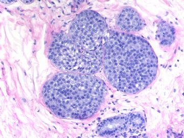
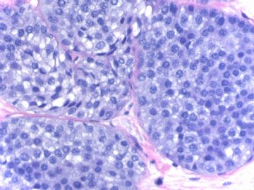
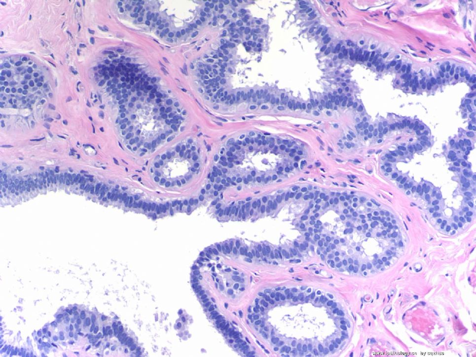
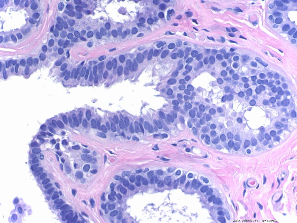
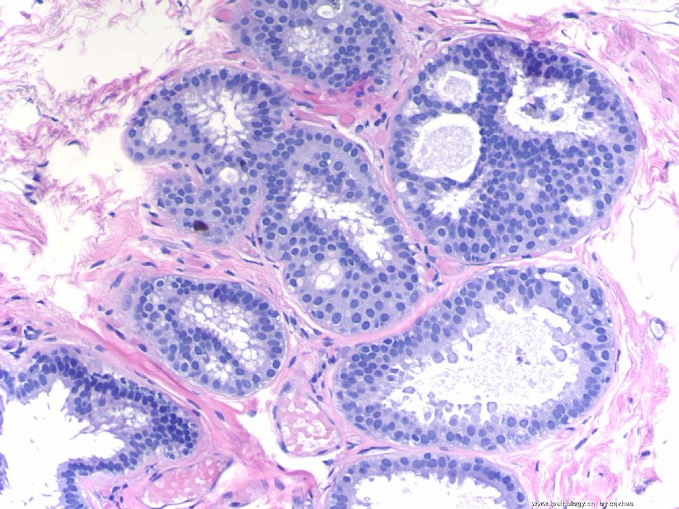
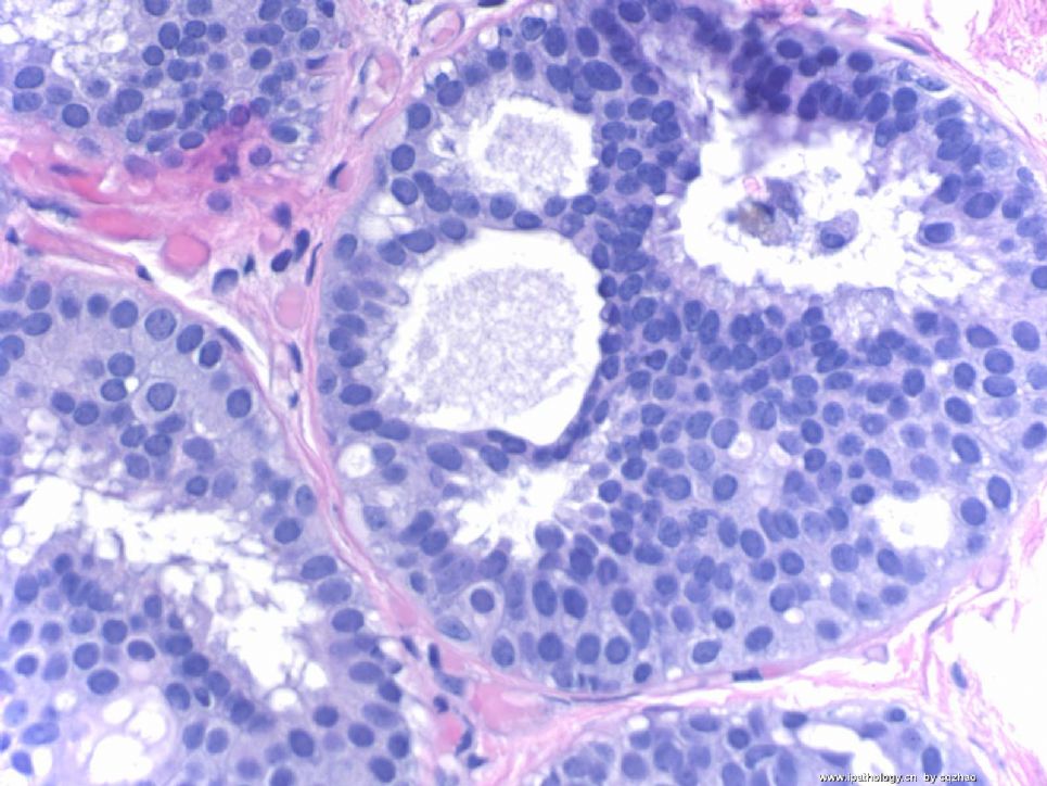
Breast core biopsy with 8 cores. There are multiple breast lesions. Try you diagnosis one by one.
Lesion 1 20x and 400x
-
本帖最后由 于 2009-03-19 23:29:00 编辑
相关帖子
- • 乳腺癌?
- • 女性 冰冻为乳腺浸润性导管癌,现切除标本,肿块旁组织
- • 女性 33岁 乳腺肿块
- • 乳腺包块
- • 乳腺两个相邻导管内的病变
- • 乳腺肿物
- • 38岁乳腺(新加HE切片)
- • 乳腺包块。33岁
- • 左乳肿块,协助诊断
- • 乳腺肿物求助
-
本帖最后由 于 2009-03-19 23:44:00 编辑
赵大夫讲的非常好,受益匪浅,译文如下:Thank every one for giving dx for this case.感谢大家给本例的病理诊断。1. Lobular carcinoma in situ (immunostains support the diagnosis).1、小叶原位癌,免疫组化支持该诊断。2 Micropapillary apocine change or metaplasia. 2、微乳头大汗腺化生或大汗腺改变。Uniform nuclei, micropapillary growth pattern. 一致的细胞核,微乳头排列。It is completely benign.完全是良性。 Papilary or micropapillary apoccrin changes are very common. 乳头状或微乳头性的大汗腺改变是非常常见的。It is part of fibrocystic changes (FCC).这是纤维囊性变的一部分(FCC)。 Generally I just call FCC and do not mention the apocrine cmetaplasia.一般情况下我们说纤维囊性变而不说大汗腺化生。 If they are ductal epithelial cells without apocrine change. 如果纤维囊性变的导管上皮细胞没有大汗腺改变,The lesions with the same cytomorphology need to be diagnosed as ADH or low grade DCIS with micropapillary pattern. 而这种改变有相同的细胞形态,应该被考虑为非典型导管增生或有微小乳头的低级别导管原位癌,This is why I pasted this photo here.这就是我为什么要贴本例的原因。3. Columinar cell change (CCC): Single layer of columnar cells lining the dilated acini with secretion. 3、柱状细胞改变(CCC):单层柱状上皮细胞衬复在有硬化扩张的腺泡上,I do not see atypia even though some nuclei are round.虽然其核圆,我没有看到异形性, It is not enough to call flat epithelial atypia even though the lining cells are not classic columinar cells.虽然内衬上皮细胞不是典型的柱状细胞,不能称之为扁平上皮非典型性。4. Intraductal papilloma.4、导管内乳头状瘤。6. Radial scar. 6、放射状疤痕,It is not a wrong diagnosis if you call sclerosing diangosis.如果称为硬化性腺病,也不错。 They are in the same categry of the lesions.这是同一范畴的病变。In our hospital if diagnosis is radial scar in breast core biopsy, breast surgeons will do an excisional biopsy. 在我们医院,粗针活检如果诊断放射状疤痕时,乳腺外科医生会做切取活检。So we are cautious for this diagnosis if no other severe lesions in the breast core.因此如果乳腺粗针活检没有其它严重病变时,我们诊断放射状疤痕非常慎重。 I think there were some studies which indicated tha radial scars increase the risk of cancers.我看到许多文献提出放射状疤痕增加了癌肿危险。 But I am really not sure it is true.但我不确定确实如此。 So if it is not a typical radial scar, I generally will not call it. 因此如果不是典型的放射状疤痕我们也不诊断。I may call sclerosing adenosis.我们称为硬化性腺病。 For this case I think it is reasonable to call radial scar.本例,有理由认为它就是放射状疤痕。OK, I have to do sth now. We will discuss the photo 5 and 7.以后会讨论图5-7.You can write your oppinion about the 5 lesions if you do not agree with me.如果谁不同意以上5种意见也可以写出自己的意见。I f you agree, we can concentrate on the other two lesions now.如果同意,这两例讨论总结至此。Thanks,cz谢谢大家。赵大夫。
Thank every one for giving dx for this case.
1. Lobular carcinoma in situ (immunostains support the diagnosis.
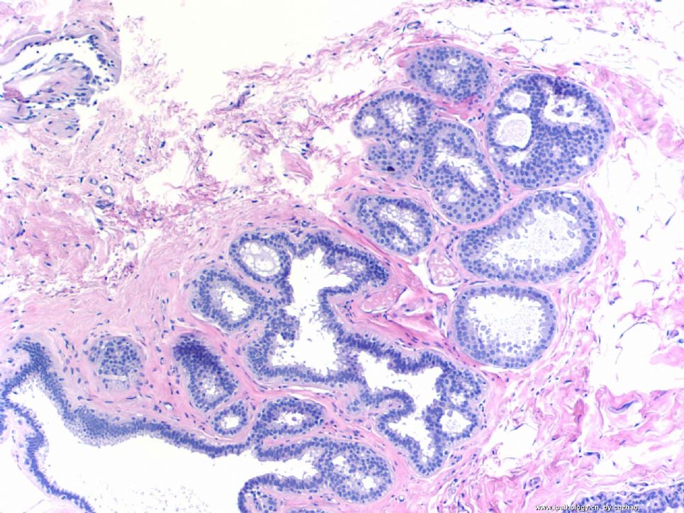
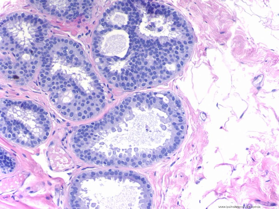
2 Micropapillary apocine change or metaplasia. Uniform nuclei, micropapillary growth pattern. It is completely benign. Papilary or micropapillary apoccrin changes are very common. It is part of fibrocystic changes (FCC). Generally I just call FCC and d not mention the apocrine cmetaplasia. If they are ductal epithelial cells without apocrine change. The lesions with the same cytomorphology need to be diagnosed as ADH or low grade DCIS with micropapillary pattern. This is why I pasted this photo here.
3. Columinar cell change (CCC): Single layer of columnar cells lining the dilated acini with secretion. I do not see atypia even though some nuclei are round. It is not enough to call flat epithelial atypia even though the lining cells are not classic columinar cells.
4. Intraductal papilloma.
6. Radial scar. It is not a wrong diagnosis if you call sclerosing diangosis. They are in the same categry of the lesions.
In our hospital if diagnosis is radial scar in breast core biopsy, breast surgeons will do an excisional biopsy. So we are cautious for this diagnosis if no other severe lesions in the breast core. I think there were some studies which indicated tha radial scars increase the risk of cancers. But I am really not sure it is true. So if it is not a typical radial scar, I generally will not call it. I may call sclerosing adenosis. For this case I think it is reasonable to call radial scar.
OK, I have to do sth now. We will discuss the photo 5 and 7.
You can write your oppinion about the 5 lesions if you do not agree with me.
I f you agree, we can concentrate on the other two lesions now.
Thanks,
cz
-
shn-821128 离线
- 帖子:277
- 粉蓝豆:3
- 经验:277
- 注册时间:2008-11-02
- 加关注 | 发消息
Quickly review all of your interpretation. Most of them are reasonable. In this topic I will concentrate for the interpretaion of FEA, columnar cell change. If you are interested you can review the text book or related articles for some photos first. Then we can have some discussion together. Dr. Stuart, J. Schnitt from Beth Israel Deaconess Medical Center, Harward Medical School, Boston, MA did a lot of study in this area. In fact he raised some terms. Frequently speaking, I am often confused by the diagnosis of FEA in my clinical practice.
Hope more people share the diagnosis about my first 7 lesions from the same patient.
Thanks,
cz
-
1. lobular neoplasia
2. nuclear grade 1 DCIS, apocrine type
3. flat epithelial atypia
4. florid ductal hyperplasia/intraductal papilloma
5. flat epithelial atypia and nuclear grade 1 DCIS
6. radial scar
7. a. columnar cell change, b.atypical coloumnar cell change, c. cribriform DCIS
1. Lobular carcinoma in situ.
2.atypical duct hyperplasia, papillary variant.
3.dilated duct.
4. papilloma.
5.columnar cell change
6.sclerosing adenosis
7.columnar cell lesions including columnar cell change, columnar cell hyperplasia,flat epithelial atypia(atypical duct hyperplasia?)
| 以下是引用luolili在2009-2-18 14:22:00的发言:
此病变有以下特征: 1 导管上皮增生,伴有轻-中度非典型增生 2 导管上皮乳头状增生,伴大汗腺化生 3 导管囊性扩张伴分泌物潴留 4 导管内乳头状瘤 5 ?是导管良性病变 6 纤维囊性乳腺病 7 腺病,导管上皮增生,伴中度非典型增生。 新手上路,望各位老师多多指教。 |
Breast pathology is complicated. We as pathologists need to recognize the cancers and all other borderline or benign lesions. There are several lesions in this breast core specimen. Please make your dx based on above orders, lesion 1, 2, 3,....
Thanks,
cz
