| 图片: | |
|---|---|
| 名称: | |
| 描述: | |
- B1581Breast lesions cqz (12) (2-16-2009)
| 姓 名: | ××× | 性别: | 年龄: | ||
| 标本名称: | |||||
| 简要病史: | |||||
| 肉眼检查: | |||||
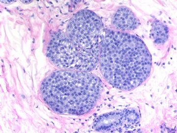
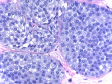
Breast core biopsy with 8 cores. There are multiple breast lesions. Try you diagnosis one by one.
Lesion 1 20x and 400x
-
本帖最后由 于 2009-03-19 23:29:00 编辑
相关帖子
- • 乳腺癌?
- • 女性 冰冻为乳腺浸润性导管癌,现切除标本,肿块旁组织
- • 女性 33岁 乳腺肿块
- • 乳腺包块
- • 乳腺两个相邻导管内的病变
- • 乳腺肿物
- • 38岁乳腺(新加HE切片)
- • 乳腺包块。33岁
- • 左乳肿块,协助诊断
- • 乳腺肿物求助
Thank Dr. Chiang' s interpretation.
Lesion 2: Apocrine metaplasia or change with micropapaillary or papillary pattern. The nuclei are very uniform, small, evenly spaced, located the botton of the cells. I do not appreciate any cytologic atypia in this case. Cystic papillary apocrine changes are commonly seen in the breast pathology specimens. Generally people will ignorred the lesion even though mild cytologic atypia is present in the apocrine change. True atypical apocrine metaplasia is rare.
-
本帖最后由 于 2009-03-19 10:28:00 编辑
Peter Rosen is an excellent breast pathologist. His Book " Rosen's Breast Pathology in the best comprehensive book in the area of breast pathology. His calssification about columnar cell lesion is different from most others. He used the term columinar cell hyperplasia, mild, moderate, and severe atypia. I really do not like his concept about CCC. It is true that intraductal carcinoma, flat pattern, is present. However, I fell reluctant to call the 5 photos Elizabeth took from Rosen's book as carcinoma based on the photos only.
-
本帖最后由 于 2009-03-14 10:40:00 编辑
Compared Rosen fig 11.11 (page 298 in the third edition) with the above lesion 3, we can appreciate the difference in term of cytology, uniform without atypia in lesion 3 and moderate cytologic atypia in Rosen's fig 11.11.
It is very good for the disagreement. I hope more pathologists join in the diacussion.
-
Fig. 11.11. Intraductal carcinoma, flat (“clinging”) micropapillary type. A: The papillary structures in this cystically dilated duct contain fractured calcifications of the ossifying type typically associated with columnar cell lesions. B: Carcinoma cells with pleomorphic nuclei and a disorderly distribution line the duct and overlie the calcification. C: The flat carcinomatous epithelium displays apical apocrine-type cytoplasmic “snouts.” D,E: Another example of flat, “clinging” intraductal carcinoma
_ from Rosen Breast Pathology. Compare with lesion 3, just for learning.
Lesion 2:大汗腺化生也存在一个谱系,良性---------恶性。该例大汗腺化生表现导管内增生,形成微乳头,细胞异型明显,核增大,核质比增加,核仁明显。但是这种改变在切除标本中未必有不同的临床意义。
Lesion 3:囊性高分泌性导管原位癌,表现导管高度扩张,腔内有分泌,被覆细胞简单,可有微乳头结构,细胞核增大,其余细胞学较温和,容易漏诊,要结合整体组织学,本例给出的图像值得怀疑和商榷。
Lesion 4:基本结构是腺病,导管有旺炽型增生,增生的导管上皮成分复杂、间质硬化,结构类似硬化性腺病,整体图像不支持导管内乳头状瘤,最多可以诊断adenosis with florid ductal hyperplasia。
Lesion7:这是一例柱状细胞变化很好的例子,显示了柱状细胞变化的形态学谱系,形态学表现延续了良性和非典型性,局灶性DCIS(需要免疫标记证实),但是这样的例子和诊断定会有争议,因为柱状细胞变化的研究还在继续
谢谢楼主提供这么好的病例。
-
Glad to see Dr. Ciang's input and diagnosis. Different interpretations were noted in lesions 2, 3, 4, and 7. Wish other share your oppinions. Also welcome Dr. Chiang to describe the reasons for the diagnoses. We can understand more by discussion. Thanks, cz
Lesion 1:lobular neoplasia,the diagnosis must be established by immunostaining for CK 34BE12.
Lesion 2:atypical ductal hyperplasia ,apocrine.
Lesion 3:cystic hypersecretory carcinoma,DCIS
Lesion 4:sclerosing adenosis with focal atypical ductal hyperplasia ,apocrine
Lesion 5:columnar cell chang,mild cytologic atypia
Lesion 6:complex sclerosing lesion
Lesion 7:columnar cell change,with severe atypia and low-grade DCIS
Fig 7 is a very good example showing the spectrum of CCC-FEA-ADH. Focal monotonous proliferation with cribriform pattern shows the features of ADH. Few glands surrounding the focus of ADH demonstrate atypical CCC or FEA. It is not easy to find the good area. Today I joined one day of breast long clourse. There are 8 excellent speakers (pathologists, oncologists, surgeons) from England, austrlia, the US talked about ADH, DCIS, FEA, bx, et al. There are many new and converoversial issue about breast pathology. There are also some difference between contries. I may find a time to review some talks with you in future.
Ok. You can mention your oppinion if you do not agree with my interpretation about this case.
Thanks, cz
-
本帖最后由 于 2009-03-12 12:14:00 编辑
Fig 5.
Right: CCC
Left: Clearly we can appreciate the difference ducts in the right from the right duct. If these are the only few atypicla ducts in a breast core biopsy, I will relactant to call FEA. If there are more ducts with similar features, I may call FEA.
CCC-FEA are a continuous lesion. The cut point is difficult.
Yes, We use the terminology of columnar cell lesion in diagnosis.
The columnar cell lesion especially flat cell atypia is related to some low grade carcinoma such as tubular carcinoma, tubulobular carcinoma etc. However, I am puzzled about FEA accompany with structure complex, should it be diagnosed as atypic duct hyperplasia or FEA, just as figures shown in lesion 7? According to the definition of FEA there should not have structure complex.
Interested in the topic very much.
Thanks!
Seem most of you agree the interpretation of F 1-4, 6. You can give your reasons if you do not agree.
F 5, 7involve columinar cell change, 平坦型上皮非典型性(FEA) and related lesions. FEA is a very controversial term. There are many issues which are not clear, such as dx criteria, managment: present in in core biopsy, in excisional bx, .in surgical margins. In fact even in the US, many general surgical pathologists do not know the term or do not know how to make the diagnois. Of cause most of surgens know nothing about FEA. We have alot of consult cases from other hospitals. Often we need to talk with primary pathologists. This is why I know a lot of general pathologists do not know the term well.
Interesting to know that our pathologists in China use this term or not. Please share your knowledge about FEA with ours.
Thanks
-
alading1999 离线
- 帖子:63
- 粉蓝豆:31
- 经验:562
- 注册时间:2009-01-17
- 加关注 | 发消息


