| 图片: | |
|---|---|
| 名称: | |
| 描述: | |
- B1581Breast lesions cqz (12) (2-16-2009)
| 姓 名: | ××× | 性别: | 年龄: | ||
| 标本名称: | |||||
| 简要病史: | |||||
| 肉眼检查: | |||||
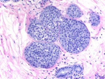
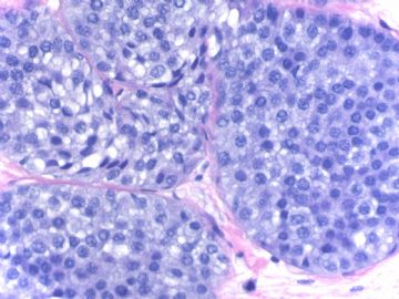
Breast core biopsy with 8 cores. There are multiple breast lesions. Try you diagnosis one by one.
Lesion 1 20x and 400x
-
本帖最后由 于 2009-03-19 23:29:00 编辑
相关帖子
- • 乳腺癌?
- • 女性 冰冻为乳腺浸润性导管癌,现切除标本,肿块旁组织
- • 女性 33岁 乳腺肿块
- • 乳腺包块
- • 乳腺两个相邻导管内的病变
- • 乳腺肿物
- • 38岁乳腺(新加HE切片)
- • 乳腺包块。33岁
- • 左乳肿块,协助诊断
- • 乳腺肿物求助
Thank every one for giving dx for this case.
1. Lobular carcinoma in situ (immunostains support the diagnosis.
2 Micropapillary apocine change or metaplasia. Uniform nuclei, micropapillary growth pattern. It is completely benign. Papilary or micropapillary apoccrin changes are very common. It is part of fibrocystic changes (FCC). Generally I just call FCC and d not mention the apocrine cmetaplasia. If they are ductal epithelial cells without apocrine change. The lesions with the same cytomorphology need to be diagnosed as ADH or low grade DCIS with micropapillary pattern. This is why I pasted this photo here.
3. Columinar cell change (CCC): Single layer of columnar cells lining the dilated acini with secretion. I do not see atypia even though some nuclei are round. It is not enough to call flat epithelial atypia even though the lining cells are not classic columinar cells.
4. Intraductal papilloma.
6. Radial scar. It is not a wrong diagnosis if you call sclerosing diangosis. They are in the same categry of the lesions.
In our hospital if diagnosis is radial scar in breast core biopsy, breast surgeons will do an excisional biopsy. So we are cautious for this diagnosis if no other severe lesions in the breast core. I think there were some studies which indicated tha radial scars increase the risk of cancers. But I am really not sure it is true. So if it is not a typical radial scar, I generally will not call it. I may call sclerosing adenosis. For this case I think it is reasonable to call radial scar.
OK, I have to do sth now. We will discuss the photo 5 and 7.
You can write your oppinion about the 5 lesions if you do not agree with me.
I f you agree, we can concentrate on the other two lesions now.
Thanks,
cz
My last four cases:
Case 1 (floor 54) columinal cell change.
Case 2-4: all these three cases were signed out by me as flat epithelial atypia (FEA).
FEA is a debatable new term. Most general pathologists in the USA do not know this lesion well. Different pathologists can have different oppinion for the same lesion.
This is fine if you do not agree above interpretation.
Ok I will complete this topic today.
Thank you for your reading or discussing these cases.
cz


