| 图片: | |
|---|---|
| 名称: | |
| 描述: | |
- B1046Breast tubular leisons, MGA and differential diagnosis (cqz 2)
| 姓 名: | ××× | 性别: | F | 年龄: | 49 |
| 标本名称: | Breast excisional biopsy (乳腺切除活检) | ||||
| 简要病史: | |||||
| 肉眼检查: | |||||
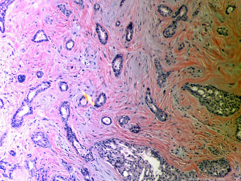
Microscopically it is a 0.8 cm lesion as photo.
Your diagnosis and differential diagnosis.
(镜下病变直径0.8cm,如图。请诊断和鉴别诊断)
名称:图1
描述:图1
-
本帖最后由 于 2010-05-16 23:38:00 编辑
相关帖子
- • 左乳肿物
- • 乳腺肿物一例
- • 腺病?癌?其他?(12楼常规,24楼免疫组化及会诊结果)
- • 左乳肿块,2X1.5cm,请会诊
- • 乳腺肿物
- • 求助:56岁女,左乳肿物,能排除小管癌吗?
- • 38岁乳腺(新加HE切片)
- • 乳腺包块。33岁
- • 乳腺肿块
- • 乳腺小管癌?
-
本帖最后由 于 2008-12-24 20:14:00 编辑
| 以下是引用天山望月在2008-12-15 21:39:00的发言:
个人感觉: 仅从细胞图片看,有时不易诊断小管癌,需与导管内乳头状瘤或癌、DCIS、低级别的浸润性导管癌、淋巴瘤等鉴别。组织切片和免疫组化有助诊断。 不知对否?请赵老师点评!谢谢! |
You are right.
Like other low grade ca, TC is very difficult to make dx in FNA cytology, especially from the benign lesions. It is important you can call atypia and require core biopsy. Cell block may be helpful, but not IHC.
-
本帖最后由 于 2008-12-15 21:31:00 编辑
You work so hard and should get some awards.
你如此勤奋,应该获奖。
Fig 1 and 2 are two classic pathology or cytopathology board photos. Whe you see them during a test, just pick the answer tubular ca immediately and go to the next question.
图1和图2是两个经典的病理学或细胞病理学样本图片。考试时看到它们时,只要立即选择小管癌即可,直接跳到下一题。
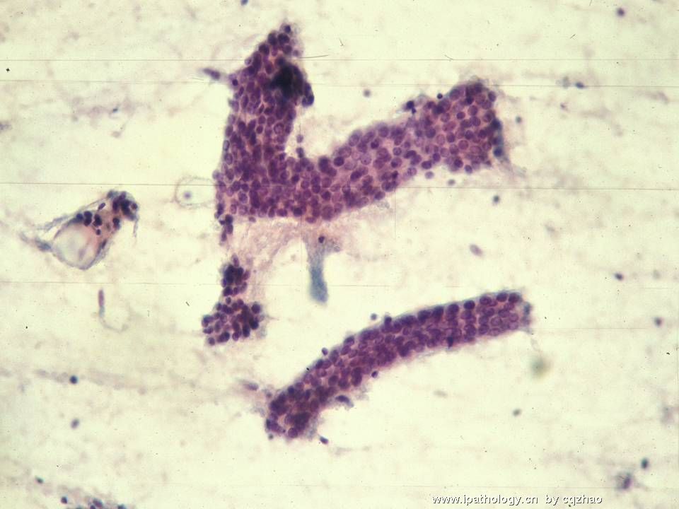
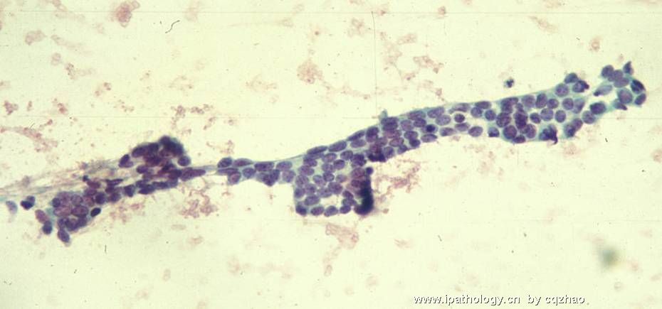
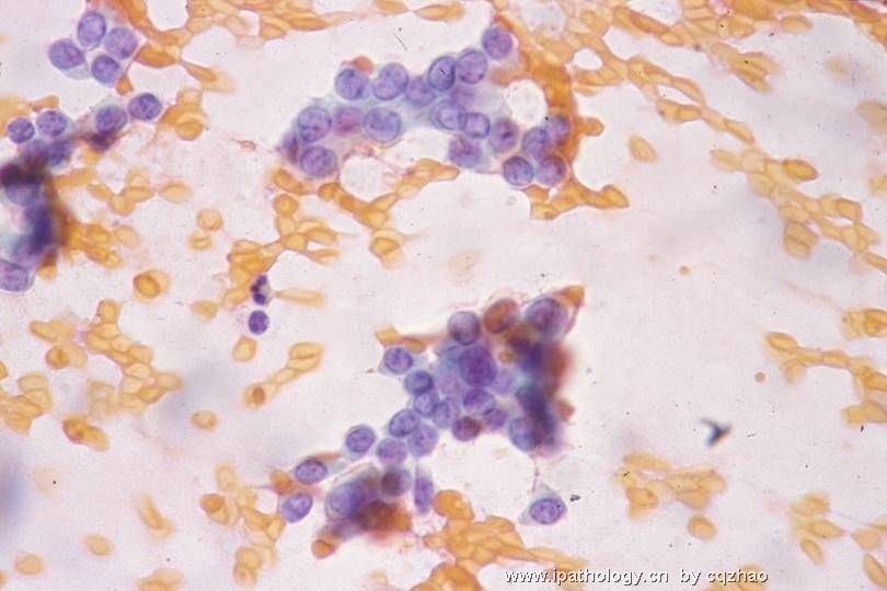
Fig 3-6 are cases of tubular ca FNA confirmed by biopsy. In true practice, you must be cautious to call tubular carcinoma for these cases. It is better to call atypical, favor... et al. Ask to do the biopsy. It is very difficult to diagnose tubular ca by FNA cytology. Find a good FNA book to read the cytopathologic features of these lesions.
图3-6是小管癌的FNA表现,由活检证实。在实际工作中,必须非常小心诊断小管癌。最好称为“不典型细胞,倾向……”等等,然后要求活检。FNA细胞学诊断小管癌非常困难。找一本FNA好书,阅读一下这些病变的细胞病理学特征。
Now you have all my photos and no more to teach you.
现在我已经全部奉献出有关小管癌的所有图片,没有更多内容用来教学了。
(abin译)

名称:图1
描述:图1

名称:图2
描述:图2
名称:图3
描述:图3
名称:图4
描述:图4
名称:图5
描述:图5
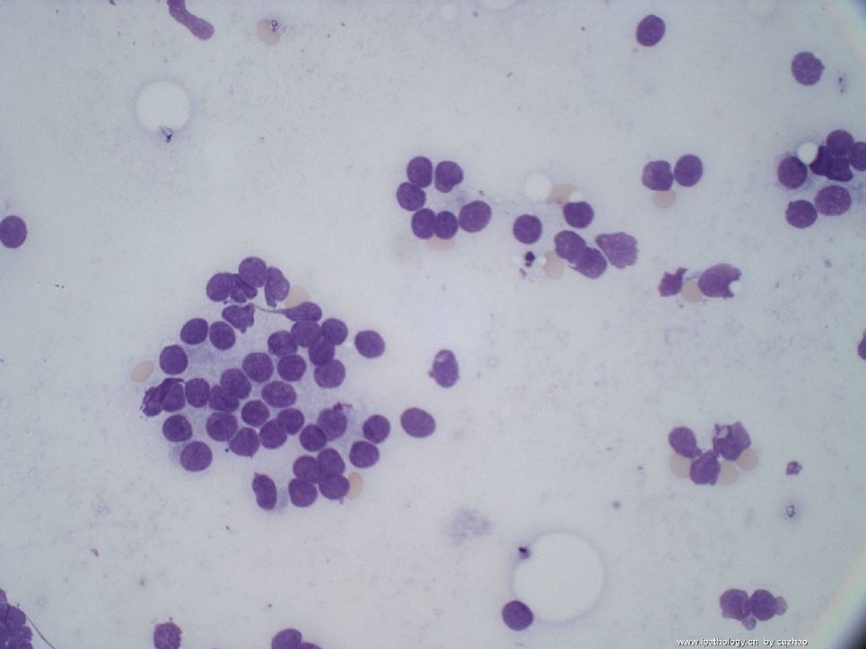
名称:图6
描述:图6
-
本帖最后由 于 2008-12-24 20:18:00 编辑
翻译如下:
为以后的教学提供一些问题,现在先选三个放这里。
欢迎写下您的答案。请注意:一个问题可以有一个以上答案。
1.小管癌细胞学,以下哪些(哪个)不是真的:
A.细胞量少
B. 小管成角,尖,僵硬,管腔开放
C. 细胞学非典型性轻微或无
D. 通常不出现双极裸核
E. 细胞簇粘附成团
F. FNA诊断敏感性较低,约50%
G. 以上均正确
2. 关于小管癌,以下哪些(哪个)不是真的:
A. 预后很好
B. 大部分 (60-70%)表现为触摸不到肿块的乳腺影像学异常
C.纯小管癌:要求 >90% 肿瘤呈现小管结构
D. 通常呈 ER/PR+, Her2-
E. 少于50% 病例伴有低级别 DCIS
G. 以上全正确
3. 哪些标记物可以区分小管癌与微腺增生
A.肌上皮标记物
B.胶原IV
C. S-100
D. CK7
E. ER

华夏病理/粉蓝医疗
为基层医院病理科提供全面解决方案,
努力让人人享有便捷准确可靠的病理诊断服务。
Made some questions for our follow teaching. Choose three and put here.
You are wellcome to write your answers. Please pay attention: One question may have more than one answers
1. Which is (are) not true about tubular carcinoma cytology
A. Hypo-cellular specimen
B. Angulated, pointed, open rigid tubules
C. Little or no cellular atypia
D. Bipolar naked nuclei usually not present
E. Cohesive clusters of cells
F. The sensitivity for the dx is lower for FNA, 50%
F. All are true.
2. Which is (are) not true about tubular carcinoma
A. Have excellent prognosis
B. Majority (60-70%) now present as nonpalpable mammographic abnormality
C. Pure TC: >90% of the tumor should exhibit tubules
D. Usually ER/PR+, Her2-
E. Less than 50% cases have associated with low grade DCIS
F. All are true.
3. Which markers are useful to distinguish tubular carcinoma from microglandular hyperplasia
A. Myoepithelial markers
B. Collagen IV
C. S-100
D. CK7
F. ER

 重新阅读本主题,不好意思
 ,第一次没审清题,再次回答51楼的问题,并期待赵老师解答:
 好可惜,奖品没拿到


