| 图片: | |
|---|---|
| 名称: | |
| 描述: | |
- B1046Breast tubular leisons, MGA and differential diagnosis (cqz 2)
| 姓 名: | ××× | 性别: | F | 年龄: | 49 |
| 标本名称: | Breast excisional biopsy (乳腺切除活检) | ||||
| 简要病史: | |||||
| 肉眼检查: | |||||
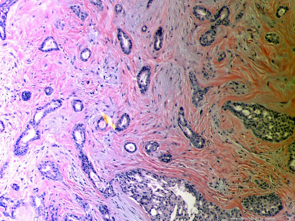
Microscopically it is a 0.8 cm lesion as photo.
Your diagnosis and differential diagnosis.
(镜下病变直径0.8cm,如图。请诊断和鉴别诊断)

名称:图1
描述:图1
标签:乳腺浸润性小管癌 硬化性腺病 微腺性腺病
-
本帖最后由 于 2010-05-16 23:38:00 编辑
相关帖子
- • 左乳肿物
- • 乳腺肿物一例
- • 腺病?癌?其他?(12楼常规,24楼免疫组化及会诊结果)
- • 左乳肿块,2X1.5cm,请会诊
- • 乳腺肿物
- • 求助:56岁女,左乳肿物,能排除小管癌吗?
- • 38岁乳腺(新加HE切片)
- • 乳腺包块。33岁
- • 乳腺肿块
- • 乳腺小管癌?
×参考诊断
1楼:SA,15楼:TC,22楼:非典型性MGA
-
本帖最后由 于 2008-10-12 22:12:00 编辑
Looks like tubular carcinoma, a well differentiated invasive ductal carcinoma. It requires >75%(?) tubular formation in neoplastic glands. Lower right hand portion of the photo shows possible low grade DCIS. The stroma probably represents desmoplastic response to those tumor glands. I talk with uncertain tone "probable, possible, looks like" because a single photomicrograph is a very limited source for a significant diagnosis, i cannnot be very sure.
abin译:
像小管癌,属于一种分化好的浸润性导管癌。它要求肿瘤性腺体中小管结构>75%(译注,这个标准有争议,有人严格要求100%。WHO认为小管结构达90%作为诊断标准可能较为妥当)。
右下可能为低级别DCIS。间质可能是促结缔组织反应。


