- 2012年第27期——尿道与阴道间隙肿块(已点评)

-
图1") 图1
图1 -
图2") 图2
图2 -
图3") 图3
图3 -
图4") 图4
图4 -
图5") 图5
图5 -
图6") 图6
图6 -
图7") 图7
图7 -
图8") 图8
图8 -
图9") 图9
图9 -
图10") 图10
图10 -
图11") 图11
图11 -
图12") 图12
图12 -
图13") 图13
图13 -
图14") 图14
图14 -
图15") 图15
图15 -
图16") 图16
图16 -
图17") 图17
图17 -
图18") 图18
图18 -
图19") 图19
图19 -
图20") 图20
图20 -
图21") 图21
图21 -
图22") 图22
图22
| 性别 | 女 | 年龄 | 49 | 临床诊断 | |
|---|---|---|---|---|---|
| 临床症状 | 阴道肿块,并无疼痛 | ||||
| 标本名称 | 尿道与阴道间隙肿块 | ||||
| 大体所见 | 淡黄色卵圆形包块1枚,2cm×1.5cm×1cm,切面亮白色,质韧。 | ||||
本例图片采用麦克奥迪MoticBA410显微镜+MoticamPro285A摄像头采集制作。
点评专家:王曦
点评专家:王曦(42楼 链接:>>点击查看<< )
获奖名单:xhyong(12楼 链接:>>点击查看<< )
标签:阴道
-
本帖最后由 冰洋 于 2012-08-08 09:50:23 编辑

知之者不如好之者,好之者不如乐之者。(语出幽梦影)

×参考诊断
上皮样平滑肌瘤


诊断:血管肌纤维母细胞瘤
依据:部位可算是会阴、阴道壁肿物。无症状。体积很小。镜下:低倍边界清楚,非侵袭性生长的,但也没有明确的假包膜(这点和平滑肌瘤有点不一样)。肿瘤细胞的分布特征构成肿块有疏松、密集区域交替。中高倍可见血管为薄壁血管,肿瘤实质细胞为上皮样、浆细胞样细胞或梭形细胞呈索状、簇状生长。但围绕血管不是很明显(这点有点和血管肌纤维母细胞瘤不一致),背景可见肥大等炎症细胞,但总体肿瘤细胞还算在薄壁血管周有规律排列生长。
IHC:CD34、Desmin、PR、ER、actin
鉴别诊断:
侵袭性血管粘液瘤:边界不清,侵袭生长,体积更大,背景更富于粘液而实质细胞更稀疏
平滑肌瘤:形态变异大,要依靠IHC
富于细胞性血管纤维瘤:玻璃样变小-中等大血管明显,肿瘤细胞排列无规律,很随意。和本例完全相反

-
本帖最后由 TK1905 于 2012-07-07 07:56:23 编辑
- Superficial, circumscribed bland lesion of external female genital tract
- Superficial cervicovaginal myofibroblastoma
- Restricted to external female genital tract
- Vagina, cervix, vulva
- Superficial circumscribed
- No capsule
- Frequent grenz zone between lesion and epithelium
- No entrapped fat
- Uniform bland stellate and spindled cells
- Oval, elongate nucleus, frequently wavy
- Even chromatin
- Small nucleolus
- 2/26 cases with a few multinucleated cells
- 4/26 cases with mild pleomorphism
- Variable patterns of cellularity
- Moderately to highly cellular overall
- Densely collagenous foci with vague fascicles
- Myxoid foci
- Lace-like or sieve-like pattern
- Cells surround nodules of collagen or myxoid stroma
- Mean age 55 years
- Only 2 of 26 cases under age 38
悲催啊!在鉴别诊断中又看见一个病:浅表性肌纤维母细胞瘤(又名浅表性宫颈阴道肌纤维母细胞瘤),觉得也很像,甚至就是它,但除了一点,即本例取材整个肿块未见阴道粘膜上皮覆盖在肿块之上,不知道手术中该肿块是否真位于阴道粘膜表浅???
浅表性肌纤维母细胞瘤:好发于阴道(占绝大多数)、宫颈、会阴,年龄绝大多数是围绝经期、绝经后,体积也很小,大多低于6cm,平均2-3cm左右。
镜下无包膜,常位于阴道的粘膜鳞状上皮下,肿块与粘膜下软组织有不清楚的交界。
血管不显著,细胞可呈中等数量到高度富于细胞(本例就是这样),细胞主要为星状细胞、梭形细胞,细胞温和,罕见核分裂,极少数例子伴有轻微的局灶的多形性。可以有局灶或部分上皮样细胞。也可以有疏松密集区交替。疏松区细胞呈蕾丝花边样或筛状、网状排列,富于细胞区可局灶密集胶原纤维伴有不甚清楚的束状排列,无陷入的脂肪细胞。
和血管肌纤维母细胞瘤主要鉴别点就是没有显著的血管,本例血管确实不是很突出,很少,不是主要病变。围绕血管生长不是很明显。
细胞形态上浅表性肌纤维母细胞瘤以星状+梭形为主,本例星状、梭形、上皮样均有;血管肌纤维母细胞瘤以上皮样细胞为主,且始终在薄壁血管周生长或成簇或条索
IHC:CD34、Vim、PR、ER、Desmin大多数例子均+,SMA部分例子表达。
Definition
Alternate/historical names
Diagnostic Criteria
Hum Pathol. 2001 Jul;32(7):715-25.
Superficial cervicovaginal myofibroblastoma: fourteen cases of a distinctive mesenchymal tumor arising from the specialized subepithelial stroma of the lower female genital tract.
Source
Department of Pathology, Northwestern University Medical School, Chicago, IL, USA.
Abstract
The clinicopathologic features and immunohistochemical profiles of 14 cases of a distinctive mesenchymal tumor that arises in the superficial lamina propria of the cervix and vagina and is histologically distinguishable from mesodermal (fibroepithelial) stromal polyp, including the cellular (pseudosarcomatous) variant, angiomyofibroblastoma, aggressive angiomyxoma, and other well-recognized lesions that occur in this location, are described. The lesions presented as a polypoid (n = 10) or nodular (n = 4) mass in the vagina (n = 12) or cervix (n = 2) of women ranging in age from 40 to 74 years (median, 58 years). The tumors were subepithelial in location, were well circumscribed, and ranged in size from 1 to 6.5 cm. (mean, 2.7 cm). Microscopically, the process was moderately to highly cellular and composed of relatively bland spindled and stellate-shaped mesenchymal cells embedded in a finely collagenous stroma that was punctuated by myxoid and edematous foci in 9 cases. The lesions characteristically had a multipatterned architecture with tumor cells focally assuming a lacelike/sievelike growth pattern in the more stroma-rich areas of the tumor and a vague fascicular growth pattern in the more cellular foci. Mitotic activity was minimal, and no atypical mitotic figures were identified. The tumors were immunoreactive (in decreasing order of relative strength) for vimentin (5 of 5 cases), estrogen (10 of 10 cases), and progesterone (10 of 10 cases) receptors, desmin (13 of 13 cases), CD34 (11 of 13 cases), alpha-smooth muscle actin (5 of 11 cases), and muscle-specific actin (2 of 8 cases). The desmin and CD34 antibodies highlighted the interconnecting, dendritic processes associated with many of the tumor cells. No immunoreactivity was detected for S100 protein, epithelial membrane antigen, or keratins. Follow-up data for 11 patients (range, 1 to 20 years; median, 4 years) showed no recurrence or metastasis after local excision. The term "superficial cervicovaginal myofibroblastoma" is proposed because it reflects the distinguishing features of this benign, relatively site-specific mesenchymal tumor. The process probably arises as a neoplastic proliferation of hormonally responsive mesenchymal cells native to the unique subepithelial stromal layer normally found through the endocervix and vulva of adult women.
Pathology. 2005 Apr;37(2):144-8.
Superficial cervico-vaginal myofibroblastoma: a report of five cases.
Source
Department of Pathology, King Edward Memorial Hospital, Subiaco, Western Australia, Australia. colin.stewart@sjog.org.au
Abstract
AIMS:
To describe the pathological and immunohistochemical features of five cases of superficial cervico-vaginal myofibroblastoma (SCVM), a recently described mesenchymal tumour affecting middle-aged and elderly females.
METHODS:
The histological features of five cases of SCVM arising in four patients were reviewed including one case which recurred locally 9 years after initial excision biopsy. All cases were immunostained using the streptavidin-biotin technique using antisera to vimentin, smooth muscle actin, desmin, S100 protein, cytokeratin, h-caldesmon, calponin, CD99, CD117 (c-kit), bcl-2, oestrogen receptor and progesterone receptor.
RESULTS:
The patients were aged from 40 to 71 years (mean 55.2 years). The tumours were situated within the vagina (four cases) and cervix (one case) and ranged from 16 to 45 mm in greatest dimension. One patient had two separate vaginal SCVM. The tumours were characterised by uniform spindle and stellate-shaped cells separated by a collagenous or myxoid stroma. No mitotic activity was identified. Characteristically the tumours were well circumscribed and separated from the surface epithelium by a rim of normal stroma. The initial and recurrent tumours in one patient were similar except for increased stromal collagen in the recurrence. All tumours were immunoreactive for vimentin, desmin, CD34, CD99, bcl-2, calponin and hormone receptors while two tumours showed focal smooth muscle actin expression. There was no expression of S100 protein, h-caldesmon, CD117 or cytokeratin.
CONCLUSIONS:
SCVM appears to be a relatively distinct lesion although there is some histological and immunophenotypical overlap with other mesenchymal tumours, particularly fibroepithelial polyp, leiomyoma and solitary fibrous tumour. As local recurrence developed 9 years after intial treatment in one patient, long-term clinical follow-up would seem appropriate.
Hum Pathol. 2012 Feb;43(2):243-53. Epub 2011 Aug 4.
Vulvovaginal myofibroblastoma: expanding the morphological and immunohistochemical spectrum. A clinicopathologic study of 10 cases.
Source
GF Ingrassia Department, Section of Anatomic Pathology, Policlinico Universitario-Vittorio Emanuele, University of Catania, 95123 Catania, Italy. g.magro@unict.it
Abstract
We analyzed the clinicopathologic features of 10 cases of vulvovaginal myofibroblastoma to widen its morphological and immunohistochemical spectrum. Most tumors (8/10 cases) were located in the vagina. The patients' age ranged from 44 to 77 years, and tumor size ranged from 0.4 to 3 cm. Histologically, 5 tumors had the characteristics of vulvovaginal myofibroblastoma. In addition, we identified 3 cases composed of spindle-shaped cells arranged in short fascicles with intervening thick collagen bands, closely reminiscent of mammary myofibroblastoma. Notably, 1 case resembled Sertoli cell tumor, sclerosing type, because of its predominant cord-like arrangement. In another case, there were highly cellular areas composed of uniform-packed, rounded cells that, at low magnification, looked like a malignant "small round blue cell tumor." A variably thick band of native connective tissue separated tumors from the overlying squamous epithelium even if, in 3 cases, tumor cells extended up to the epithelium. In 7 cases, a variable number of vessels showed perivascular hyalinization. Only rare mitotic figures were identified. All tumors were diffusely positive for vimentin, desmin, and CD99. A variable staining intensity was observed for CD34, Bcl-2, B-cell lymphoma 2 (Bcl-2) CD10, estrogen receptor, and progesterone receptor in most cases, but none expressed α-smooth muscle actin. We emphasize that vulvovaginal myofibroblastoma encompasses a morphological spectrum wider than previously described. The overlapping morphological and immunohistochemical features of vulvovaginal and mammary myofibroblastomas led us to speculate that these are related entities with morphological variations on a common basic theme likely dependent on anatomical location.
DISCUSSION
A wide variety of mesenchymal lesions occur in the lower female genital tract.5,6 Broadly, these mesenchymal lesions can be separated into two groups. The first group includes several well-characterized tumors that show a marked tendency to occur in the lower female genital tract, such as aggressive angiomyxoma, angiomyo- fibroblastoma, and cellular angiofibroma.7-9 These tumors can also be called as relatively site-specific. The second group embraces a wide range of heterogeneous lesions that frequently occur in this region, but arise in other anatomic sites as well, with examples as fibroepithelial stromal polyp, superficial angiomyxoma and leiomyoma.
SCVM represents a new member of the first group. SCVM appears underrecognized. To our knowledge, only 31 cases of SCVM have been reported so far in the literature.1-4 The current four cases raise the number to 35. These 35 cases occurred in 34 patients ranging in age from 23 to 80 years with mean of 57 years. To date, 32 patients (94%) were in their peri- or post-menopausal years. Eleven (32%) patients had received Tamoxifen or hormone- replacement therapy. Clinically, the most common presentation was an asymptomatic polypoid nodule or mass, with cysts or polyps being the most preoperative diagnosis. Thirty patients presented with solitary nodular mass, whereas 4 patients had two separated lesions. Thirty tumors were located in the vagina or vaginal fornix, 3 in the cervix and 2 in the vulva. Because the tumor also occurred in the vulva, Ganesan et al2 suggested the term superficial myofibroblastoma of the lower female genital tract to replace SCVM.
Overall, most SCVM were described as polyps or had a polypoid outline, measuring 1.0 cm to 8.0 cm (mean, 2.5 cm) in diameter. Histologically, SCVM was characterized by its superficial subepithelial location and uniform spindle and stellate cells arranged in diverse growth patterns within a myxoid to collagenous stroma. Tumor cells in SCVM typically expressed vimentin, desmin, CD34, ER and PR, with desmin and CD34 accentuating the dendritic processes of the cytoplasm, whereas actins was consistently negative.
Due to some histological and immunohistochemical overlapping features, SCVM can be confused with other mesenchymal lesions of the lower female genital tract. The principal differential diagnosis is FSP, especially the cellular variant.9 As described in the original paper, 4 cases had been primarily diagnosed as FSP. The younger population of FSP, absence of distinct Grenz zone in FSP, lack of heterogeneous cellularity and diverse growth patterns, are crucial features to distinguish SCVM from FSP.9 The other tumor enters the differential diagnosis is angiomyofibroblastoma (AMF). AMF is also characterized by alternating hypercellular and hypocellular areas. However, unlike SCVM, AMF contains abundant small to medium sized blood vessels. The tumor cells in AMF also have a propensity to grow around the vessels. Furthermore, SCVM is frequently negative for α-smooth muscle actin. In contrast, AMF is usually positive for actins.8 Cellular angiofibroma (CA) is another site-specific mesenchymal tumor that should be considered in the differential diagnosis. CA usually involves the vulva and the inguinoscrotal region. CA differs from SCVM by its numerous small- to medium-sized thick-walled vessels and negative stainging for desmin. Other lesions that may be confused with SCVM include leiomyoma, solitary fibrous tumor (SFT) and mammary-type myofibroblastoma.10 Vulvovaginal leiomyoma often shows a hyalinized or myxoid stroma, so called myxohyaline pattern. In addition to desmin, ER and PR, the tumor cells in leiomyoma are also positive for actins. SFT is another tumor which may display alternating cellularity. In contrast to SCVM, SFT is typically positive for CD34 and negative for desmin. Mammary-type myofibroblastoma is a spindle cell tumor with morphological and immunophenotypic features identical to myofibroblastoma of breast. Mammary-type myofibroblastoma shows a predilection for the inguinal area of older men. Only one case has been reported in the vaginal wall.11 In our opinion, this case is possibly a lesion of SCVM.
The pathogenesis of SCVM is not well understood. However, nearly one third of SCVM patients had a history of Tamoxifen or hormone-replacement therapy, suggesting that hormones may possibly have a role in the development of the tumor. It has been postulated that the subepithelial mesenchymal cells of the lower female genital tract, which are normally estrogen and progesterone positive, tend to be the origin of most site-specific mesenchymal tumors that arising in this site.1
Follow-up information was available in 30 cases of SCVM. Twenty-nine cases pursued a benign outcome with no evidence of recurrence or metastasis. Only one patient experienced local recurrence 9 years after initial excision biopsy.3 As there is limited potential for local recurrence, complete local excision is curable for SCVM. Nevertheless, long-term follow-up is recommended.
In conclusion, SCVM represents a distinctive entity which shows a marked predilection for the superficial aspect of the lower female genital tract, predominantly the vagina. Hormones may be involved in the growth of the tumor. Although this new entity is of little relevance to the gynecologist for therapeutic decisions, awareness of its unique clinical and histological features is helpful for the pathologists at differential diagnosis.


MESENCHYMAL LESIONS OF THE LOWER GENITAL TRACT
Deep (aggressive) angiomyxoma ;Fibroepithelial stromal polyp ; Superficial angiomyxoma
Angiomyofibroblastoma ; Cellular angiofibroma ; Extramammary myofibroblastoma; Prepubertal vulval fibroma
本例我考虑是一上皮样型平滑肌瘤,具体阐述鉴别,待续
-
本帖最后由 xhyong 于 2012-07-07 14:12:32 编辑

也不想抄书了,本例几个特点:肿瘤发生在会阴生殖部位,此部位的梭形细胞病变抄书一大堆,大家也能查到
另一个特点:细胞呈条索样,上皮样
呈条索样、上皮样的软组织肿瘤大家可能有了解很多:比如上皮样平滑肌瘤 上皮样血管内皮瘤,上皮样血管肉瘤,间皮瘤,副脊索瘤/肌上皮瘤 粘液样软骨肉瘤 部分GIST
本例除了上皮样细胞外,还可以见到梭形细胞,13、14可以见到两者共存,还略有过渡
同时有广义的两种分化的软组织肿瘤:间皮瘤,血管内皮瘤,GIST ,滑膜肉瘤,MPNST,平滑肌肿瘤
不同的同行在诊断一个肿瘤时,特别是疑难肿瘤,都有自己的观点或落脚点,主要看自己更在乎哪一种改变,哪一种改变对你的印象最深
此例给我的印象:女性会阴部位,上皮样条索,胞浆红染,并没有纯梭形细胞,或纯上皮样细胞,没有显著的厚壁薄壁血管改变,没有所谓的围绕血管的上皮样细胞簇,没有典型的粘液改变,没有典型的绳索样胶原纤维
故我考虑是一个上皮样平滑肌瘤
这种改变:在骨外粘液样软骨肉瘤,上皮样血管内皮瘤,甚至副脊索瘤中有类似表现,也如前所述,自己更在意什么改变,我可以认为没有典型的腺管样结构,典型的三个一,粘液样背景的条索样略淡红色细胞,还有自己的feeling,觉得虽然 部分区域想粘液软骨肉瘤,但是结合部位和性别,可能会pass掉,虽然两者形态也符合副脊索瘤/肌上皮瘤,但是也是考虑位置性别等原因,也不首选
鉴别诊断:免疫组化可以写很多,前面很多同仁皆列出了,我认为本例CD34 ER PR SMA caldesmon HHF35 S-100在诊断和鉴别诊断中有帮助
最后附带传一张截图,也许不甚相关,大家共同学习提高

-
本帖最后由 红胜火 于 2012-07-07 14:35:02 编辑
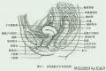 图1
图1 图1
图1 图1
图1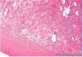 图2
图2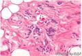 图3
图3 图1
图1 图2
图2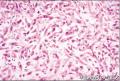 图1
图1 图1
图1 图2
图2 图1
图1 图2
图2
刚忙完,发现又一例上线了,而且很多高手已经出招
这一例我仔细学习了高手们的意见后,又好好看了图片,主要的体会如下:
女性,发生部位比较特殊,阴道与尿道间隙。这个部位在什么地方呢?
从图上看出,女性尿道位于阴道前方,与阴道壁紧密相邻,所以发生于阴道与尿道间隙的肿瘤有3种可能,1.部位相关性软组织肿瘤,主要是与阴道紧密相关,2.非部位相关性软组织肿瘤,3.侵袭性肿瘤结节(来自尿道或阴道恶性肿瘤)。
再来看标本,最大径2cm,比较小啊,临床大夫应该不会搞错解剖部位,虽然时常见他们搞错,尤其是肿物很大时, 。所以这个肿物的部位应该比较深,不会很表浅。
再看镜下:
1.界限清楚的结节,局部可见纤维性假包膜,但大多不见包膜。
2.镜下特征性形态是细胞呈条索状、网织状排列、花边状排列,瘤细胞圆形、多边形、梭形,有的细胞可见胞浆内空泡,有的空泡内含红细胞。染色质细腻,可见小核仁,核分裂像不容易看到。间质呈水肿、粘液样改变,局部硬化。低倍下略有分叶状改变。血管特点是主要是一些薄壁小血管,同时可见中等大小的厚壁血管,扩张。
综合以上,首先考虑的病是软组织肌上皮瘤。上皮样细胞、网格状排列,间质粘液变、硬化,这些提示可能是肌上皮瘤。免疫组化CK、S-100阳性应该可以明确。
免疫组化:CK、EMA、S-100、CD31、CD34、SMA、desmin、h-Caldesmon、ER、PR、。
本例需要鉴别的疾病很多,根据对发病部位的分析,主要有以下鉴别诊断:
一、女性生殖道部位特异性软组织肿瘤。很多了,主要如下表:
这一例形态最难与血管肌纤维母细胞瘤鉴别。因为后者常常也会出现上皮样细胞形态,聚集于血管周围,并形成细胞巢或细胞索。没有首先考虑这个病的原因是(1)本例肿瘤内血管还是比较少的,(2)围绕血管生长的模式还是不清晰,(3)间质胶原不多,细胞疏松区与致密区交替不明显。但仔细观察可见少量梭形成束状排列的细胞,所以这个诊断还是不能轻易排除。
第二个需要鉴别的部位特殊性病变就是浅表性宫颈阴道肌纤维母细胞瘤(SCVM)。这个单纯从形态上也是很难鉴别,因为它也会表现为上皮样细胞、网格状、条索状、花边状排列,尤其是本例具有的梭形成束细胞,让我实在难以割舍SCVM。可是本例显示它的常见大体改变为息肉样,至少是位于粘膜下,与粘膜有一隔离带,周边粘液水肿样,形态似本例改变,中央或深部则为密集的具有很多胶原纤维的梭形细胞区,很少出现分叶状改变。
第三个需要鉴别的是上皮样平滑肌瘤。平滑肌瘤形态变化多端,尤其是发生于女性生殖道时,而且发病率又极高,拉上它绝对不为过。免疫组化SMA、desmin、h-Caldesmon可以帮助诊断。
免疫组化如果排除了肌上皮瘤的诊断,以及下面的鉴别诊断,在血管肌纤维母细胞瘤和SCVM之间选择的话,还真的是很难,需要进一步详细了解临床特征。
二、非部位特殊性软组织肿瘤。
主要考虑与下面3个病鉴别:
1.上皮样血管内皮瘤,形态与本例形态很像,但它常常呈浸润性生长。本例界限很是比较清楚。
2.非骨化性纤维粘液样肿瘤。骨化性纤维粘液样肿瘤多有一层外周骨壳,肿瘤略呈分叶状,细胞呈短条索状、网格状排列,可以有梭形束状细胞区,当没有骨壳时,有的作者将其称为非骨化性纤维粘液样肿瘤。本例形态与该病很像,而且界限较清这一点也积极提示可能是这个病。但总感觉纤维样背景不明显。也是难以割舍的一个诊断 。
。
3.另外有个呈网状排列的软组织肿瘤,即网织状神经束膜瘤。多有纤维性假包膜,镜下出现特征性的网格状排列,间质粘液样,细胞梭形,核卵圆形。但一般有密集梭形区,呈席纹状排列。本例缺乏。免疫组化EMA阳性可以帮助我们。
总之,本例很难,与这个病很像,又和那个病很像,实在难以抉择,最后选择肌上皮瘤,主要是感觉纤细的纤维样背景,一般会出现在以上鉴别诊断中的,本例不是很明显。
最后还是感谢城北老师提供 了这么一个好病例,让我对软组织的一些病又学习了一遍,不管最终诊断如何,我感到收获大大的!

您们真厉害,佩服啊,思路好清晰,还那么详尽,不像我基本上是“搬书” ,以上各位详尽分析的老师的思路以及意见本人完全赞同。我主要还是部位上考虑,仅凭形态考虑的诚如楼上的96298、xhyong、红胜火所叙述的那样那些肿瘤都有类似形态,很多有都有形态重叠,部位在诊断上也不是绝对的指标,只不过是发病率高些罢了,一切皆有可能,到底是神马不到最后更明确的证据真不好说!
看到您们的精彩分析还图文并茂,我顿时成了您们的粉丝,对您们的敬仰有如……(此处省去500字)
再次感谢城北楼主送来了如此精彩病例,我的诊断虽然可能不正确但要搞清楚还是花了时间学习的,比如浅表性肌纤维母细胞瘤/浅表性宫颈阴道肌纤维母细胞我借着这次机会才学习它,不然我还从未听说呢
昨天我想了又想之后,以为自己是第一个把血管肌纤维母细胞瘤提出来的,窃喜,一发贴,居然TK1905这位大哥抢了沙发,居然英雄所见略同,虽然我不是英雄,但跟TK1905站在同一角度看一个病变,心里还是有英雄感的,毕竟自己才拿执医资格症三个月,呵呵。我当时翻阅了教学版块王坚老师讲的好发于女性生殖系统的软组织肿瘤这个课件,同时也考虑了浅表性肌纤维母细胞瘤,但是部位与阴道粘膜的关系不明确,不敢考虑。当然对有些细胞的形态也存在一些疑问,王坚老师的课件图上血管肌纤维母细胞肿瘤围绕血管的瘤细胞形态相当明显,此图不明显,本着临床印象为先的原则,我考虑了血管肌纤维母细胞瘤。今天再次看了红胜火,xhyong,TK1905,96298等大侠的贴子,忽觉上皮样平滑肌瘤也是一个好诊断,同时被你们宽阔的思维所折服,你们的旁征博引激励一个后辈不断上进,很希望大侠们分享一下看图片后翻书与查PUBMED的经验,查资料的方法比做一个具体诊断更重要,就像软组织肿瘤确定良恶性比具体诊断更重要。
- 我思故我在