


| 图片: | |
|---|---|
| 名称: | |
| 描述: | |
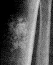
- B2238纵隔多结节性肿物,期待您的高见!
| 以下是引用XLJin8在2010-12-30 10:21:00的发言:
1、“去分化”的概念1971年由Ddhlin和Beabout首先用于软骨肉瘤(见文献1)。它指低级别软骨肉瘤(1级或2级)转变为高级别肉瘤, 后者最常为恶性纤维组织细胞瘤或骨肉瘤。此后,去分化的概念被广泛运用于许多低度恶性的肉瘤转变为高度恶性的肉瘤, 包括高分化脂肪肉瘤、平滑肌肉瘤、和低度恶性皮质旁骨肉瘤,脊索瘤等。并被用于癌(如甲状腺去分化乳头状癌) 2、由于在“去分化”后, 高级别肉瘤的形态学大多数表现为恶性纤维组织细胞瘤(MFH), 因此MFH被认为是低级别肉瘤的 “最后的共同之路” (malignant fibrous histiocytoma as a final common pathway)。 3、要注意“ 去分化”(dedifferentiation) 定义明显区别于“逆分化” (retrodifferentiation), 逆分化是指某一种分化成熟状态的细胞直接逆转为其分化成熟前状态。 4、去分化脂肪肉瘤中的非脂肪性高度恶性肉瘤成分(MFH)是否真的不再具有脂肪细胞的表型?以前是根据形态学进行判断,未被遗传学研究认证。最新研究发现并不完全如此(见文献2:Conclusion: dedifferentiated LPS can show lipoblastic differentiation in the high-grade component, resulting in areas indistinguishable from pleomorphic LPS. The available clinical and molecular data support the notion of "homologous" lipoblastic differentiation in dedifferentiated LPS, rather than mixed-type LPS.)。 5、要诊断“去分化”,必须要有高分化肉瘤的形态学依据,否则何从讲“去分化”?如何寻找:1)原发肿瘤或复发瘤中同时存在低级别肉瘤和高级别非同源性肉瘤二种成分;2)以前有高分化肉瘤的组织学依据,复发瘤为非同源性高级别肉瘤。 6、对于不同组织学类型的高分化肉瘤,诊断“去分化”的标准不同。再次复习文献发现去分化并非Dahlin 最早用于“去分化皮质旁骨肉瘤”(文献3),而是Dahlin用于“去分化软骨肉瘤”。现在看来Dahlin定义的低度恶性骨肿瘤的去分化与“去分化脂肪肉瘤”的定义不完全相同。因此,诊断“去分化”要根据具体的肉瘤类型,有的需要“转变”,有的并不需要“转变”。 1)Dahlin DC Beabout JW: Dedifferentiated chondrosarcoma. Cancer 1971;28:461-466. 2)Mariño-Enríquez A, Fletcher CD, Dal Cin P, Hornick JL. Dedifferentiated liposarcoma with "homologous" lipoblastic (pleomorphic liposarcoma-like) differentiation: clinicopathologic and molecular analysis of a series suggesting revised diagnostic criteria. Am J Surg Pathol. 2010 Aug;34(8):1122-31. 3)Wold LE, Unni KK, Beabout JW, Sim FH, Dahlin DC. 感谢谢您的提问!以上回答为个人理解,仅供参考。 |
谢谢Jin老师认真、详细的讲解。
-
本帖最后由 于 2011-01-02 17:28:00 编辑
| 以下是引用lfp在2010-12-24 22:56:00的发言:
XLJin8老师:“最初Dahlin提出的“去分化”的概念是指低度恶性的皮质旁骨肉瘤转变为高度恶性的骨肉瘤或纤维肉瘤/MFH” 请问:诊断去分化性肉瘤是否需要强调“转变”?如果是,如何寻找“转变”证据? |
1、“去分化”的概念1971年由Ddhlin和Beabout首先用于软骨肉瘤(见文献1)。它指低级别软骨肉瘤(1级或2级)转变为高级别肉瘤, 后者最常为恶性纤维组织细胞瘤或骨肉瘤。此后,去分化的概念被广泛运用于许多低度恶性的肉瘤转变为高度恶性的肉瘤, 包括高分化脂肪肉瘤、平滑肌肉瘤、和低度恶性皮质旁骨肉瘤,脊索瘤等。并被用于癌(如甲状腺去分化乳头状癌)
2、由于在“去分化”后, 高级别肉瘤的形态学大多数表现为恶性纤维组织细胞瘤(MFH), 因此MFH被认为是低级别肉瘤的 “最后的共同之路” (malignant fibrous histiocytoma as a final common pathway)。
3、要注意“ 去分化”(dedifferentiation) 定义明显区别于“逆分化” (retrodifferentiation), 逆分化是指某一种分化成熟状态的细胞直接逆转为其分化成熟前状态。
4、去分化脂肪肉瘤中的非脂肪性高度恶性肉瘤成分(MFH)是否真的不再具有脂肪细胞的表型?以前是根据形态学进行判断,未被遗传学研究认证。最新研究发现并不完全如此(见文献2:Conclusion: dedifferentiated LPS can show lipoblastic differentiation in the high-grade component, resulting in areas indistinguishable from pleomorphic LPS. The available clinical and molecular data support the notion of "homologous" lipoblastic differentiation in dedifferentiated LPS, rather than mixed-type LPS.)。
5、要诊断“去分化”,必须要有高分化肉瘤的形态学依据,否则何从讲“去分化”?如何寻找:1)原发肿瘤或复发瘤中同时存在低级别肉瘤和高级别非同源性肉瘤二种成分;2)以前有高分化肉瘤的组织学依据,复发瘤为非同源性高级别肉瘤。
6、对于不同组织学类型的高分化肉瘤,诊断“去分化”的标准不同。再次复习文献发现去分化并非Dahlin 最早用于“去分化皮质旁骨肉瘤”(文献3),而是Dahlin用于“去分化软骨肉瘤”。现在看来Dahlin定义的低度恶性骨肿瘤的去分化与“去分化脂肪肉瘤”的定义不完全相同。因此,诊断“去分化”要根据具体的肉瘤类型,有的需要“转变”,有的并不需要“转变”。
1)Dahlin DC Beabout JW: Dedifferentiated chondrosarcoma. Cancer 1971;28:461-466.
2)Mariño-Enríquez A, Fletcher CD, Dal Cin P, Hornick JL.
Dedifferentiated liposarcoma with "homologous" lipoblastic (pleomorphic liposarcoma-like) differentiation: clinicopathologic and molecular analysis of a series suggesting revised diagnostic criteria. Am J Surg Pathol. 2010 Aug;34(8):1122-31.
3)Wold LE, Unni KK, Beabout JW, Sim FH, Dahlin DC.
Dedifferentiated parosteal osteosarcoma. J Bone Joint Surg Am. 1984 Jan;66(1):53-9.
Parosteal osteosarcoma is an uncommon malignant tumor of bone, and in a review of Mayo Clinic records we identified eleven cases of so-called dedifferentiated parosteal osteosarcoma. Ten of the eleven patients had had a long history of treatment for multiple recurrences of the tumor as a low-grade parosteal
osteosarcoma and then for a definite recurrence as a high-grade undifferentiated osteosarcoma. The prognosis in this group of patients was similar to that in patients with conventional osteosarcoma.
感谢您的提问!以上回答为个人理解,仅供参考。

- xljin8
|
非常好的病例,根据形态学和IHC标记结果。前7张图为高分化脂肪肉瘤,后5张图是脂肪+平滑肌肿瘤, 二者应该以一元论解释。 如何诊断? 1)脂肪肉瘤,去分化型,低度恶性 2)去分化脂肪肉瘤(去分化成分平滑肌肉瘤)? 3)高分化脂肪肉瘤伴有低级别去分化(Well differentiated liposarcoma with low-grade dedifferentiation)? 4)高分化脂肪肉瘤伴平滑肌分化(脂肪平滑肌肉瘤 Lipoleiomyosarcoma)? 5)高分化脂肪肉瘤伴有微小去分化(高度恶性肉瘤,面积<1cm, Well differentiated liposarcoma with minimal dedifferentiation) |
-
wangzhe0439 离线
- 帖子:12
- 粉蓝豆:1
- 经验:12
- 注册时间:2010-03-02
- 加关注 | 发消息
-
本帖最后由 于 2010-12-22 21:26:00 编辑
| 以下是引用sanxia在2010-9-3 18:46:00的发言: 谢谢金主任,学习了! 不过,WHO和ACKERMAN外科病理学上都说“去分化成分可以是低级别的”,不知该如何理解?谢谢! |
非常好的问题! 试做解答:
1) 对于脂肪源性恶性肿瘤研究发展比较快的阶段是在1990年后。到2000左右基本分类已大致确定。(1)不典型脂肪肿瘤(主要位于浅表)/高分化脂肪肉瘤(体腔)。高分化脂肪肉瘤又细分为:脂肪瘤样脂肪肉瘤、炎症型脂肪肉瘤、硬化型脂肪肉瘤、去分化脂肪肉瘤。(2)黏液脂肪肉瘤/圆形细胞脂肪肉瘤。(3)多形性脂肪肉瘤。
2) ‘不典型’脂肪肿瘤的概念引入是为了避免对形态学为高分化脂肪肉瘤,但肿瘤发生在躯干和四肢浅表部位,往往临床发现早,手术易于完整切除、复发率低的这些病例治疗过度。就像把发生在皮下浅表组织的MFH称为“不典型黄色纤维瘤”一样。
3) 最初Dahlin提出的“去分化”的概念是指低度恶性的皮质旁骨肉瘤转变为高度恶性的骨肉瘤或纤维肉瘤/MFH, 原本无“异源性”要求。1979 Evan提出去分化脂肪肉瘤的概念, 并提出“非脂肪源性”和“高级别”二项要求。
4) 交界性或低度恶性肿瘤“去分化”后,一般生物学行为转变为高度恶性。但是,其恶性程度还要取决于“去分化区域“的大小和恶性度。
5) 文献中低度恶性去分化脂肪肉瘤的文章是强调“另一种肉瘤成分”的恶性程度低,区别于低度恶性脂肪肉瘤去分化后出现高度恶性转变。
6) 文献中低级别非脂肪性肉瘤成分主要为2种类型;a.平滑肌肉瘤[1];b.骨肉瘤[2]。
7) 在高分化脂肪肉瘤中出现梭形细胞[3]、平滑肌、软骨、骨组织时, 要根据其形态学特征来确定其良恶性,恶性程度为为低级别还是高级别。个人认为如果高分化脂肪肉瘤中出现低级别异源性肉瘤成分,如高分化脂肪肉瘤伴有部分高分化平滑肌肉瘤成分,高分化骨肉瘤,不提倡诊断为去分化脂肪肉瘤。真如文献1-3中指出的那样。
8)如果是原发肿瘤出现高分化脂肪肉瘤和另外一种高分化肉瘤,也可认为是‘恶性间叶瘤’。当然,有少数去分化脂肪肉瘤在首次手术标本就有高分化脂肪肉瘤和界限明显的高度恶性肉瘤区域。如果异源性成分是出现在复发性高分化脂肪肉瘤中,它可能提示肿瘤分化基因不稳定的显性化或出现了继发性分子事件。
以上纯属个人观点,仅供参考,错误之处请指正。 谢谢!
1) Folpe AL, Weiss SW.
Lipoleiomyosarcoma (well-differentiated liposarcoma with leiomyosarcomatous differentiation): a clinicopathologic study of nine cases including one with dedifferentiation. Am J Surg Pathol. 2002;26:742-9.
Leiomyosarcomatous (LMS) differentiation is a rare event in liposarcoma (LPS) and may consist of either well-differentiated liposarcoma (WDL) with an intrinsic smooth muscle component, so-called "lipoleiomyosarcoma," (L-LMS) or dedifferentiated liposarcoma having smooth muscle differentiation in the dedifferentiated zones. The latter are high-grade sarcomas, whereas the behavior of the former group is uncertain. Specifically, it is not clear whether the presence of LMS negatively affects the prognosis. We present our experience with
nine cases, the largest to date. The patients (seven male, two female) ranged in age from 42 to 65 years (mean 54 years). The tumors were usually large (2 to >40 cm [mean 17 cm]) tumors in the retroperitoneum (two cases), paratesticular-inguinal region (three cases), mediastinum (one case), lung (one case), abdomen (one case), and popliteal fossa (one case). The nine cases qualified as L-LMS and showed typical WDL with a multifocal, gradual transition into smooth muscle areas. The latter areas accounted for a variable portion of the lesions (range 5-90%) and were of low cellularity, mild to moderate nuclear atypia, and low mitotic activity. These areas seemed to arise from or blend with the smooth muscle in the walls of large vessels within the tumor. One case showed areas of dedifferentiation consisting of actin and desmin-negative, high-grade sarcoma. Follow-up in seven cases (range 26-312 months; mean 119 months) showed multiple local recurrences in seven patients and no metastases. Three patients are currently without evidence of disease (follow-up duration 26-312 months; mean 144 months) and four patients are alive with progressive disease (follow-up duration 60-132 months; mean 99 months). Our study suggests that L-LMS is a dual lineage sarcoma as evidenced by the fact that the smooth muscle component is often multifocal, not necessarily found in close association with the atypical changes in fat, and seemingly originates from atypical ("in situ") changes in the vessel wall. The LMS component, which is typically low grade, does not adversely affect the overall behavior of the tumor, which is similar to that of conventional WDL. LMS in L-LMS should not be misconstrued as evidence of low-grade dedifferentiation, a phenomenon that identifies a more unstable and potentially metastasizing lesion.
2. Yoshida A, Ushiku T, Motoi T, Shibata T, Fukayama M, Tsuda H.
Well-differentiated liposarcoma with low-grade osteosarcomatous component: an underrecognized variant.
Am J Surg Pathol. 2010;34:1361-6.
Mature bone formation in well-differentiated liposarcoma and dedifferentiated liposarcoma has been described as a reactive or "metaplastic" change in most reports, and its neoplastic nature has not been widely appreciated. We herein describe 9 cases of well-differentiated/dedifferentiated liposarcoma with distinct areas of fibroosseous tissue histologically indistinguishable from low-grade osteosarcomas, that is, parosteal osteosarcoma or low-grade central osteosarcoma. The tumors affected middle-aged to elderly patients, and occurred in the retroperitoneum and deep soft tissue of the extremities without connection to the skeletal system. Grossly, all the tumors showed biphasic appearance with lipogenic and osteogenic area, the latter representing 5% to 50% of the total tumor volume. Histologically, the lipogenic component exhibited typical histology of well-differentiated liposarcoma, whereas the osteogenic area consisted of fibroosseous tissue with numerous mature neoplastic bone trabeculae largely lacking osteoblastic rimming, with intervening fascicles of spindle cell proliferation showing low nuclear grade. All samples were positive for MDM2 and/or CDK4 on immunohistochemical analysis; the antibodies stained many osteocytes, indicating that the bone is neoplastic rather than reactive. Three cases showed high-grade osteosarcomatous transformation juxtaposed to the low-grade osteosarcomatous component, reminiscent of the "dedifferentiation" phenomenon of skeletal low-grade osteosarcoma. Follow-up revealed local recurrence in 4 cases, but no distant metastases were documented. Recognition of this earlier underappreciated subtype of well differentiated/dedifferentiated liposarcoma is important, because the fibroosseous component may seem so bland that it may be confused with benign metaplasia such as myositis ossificans, or conversely, the lipomatous component may be inconspicuous that it may be dismissed as normal fat, and such misinterpretation may potentially result in suboptimal treatment.
3) Dei Tos AP, Mentzel T, Newman PL, Fletcher CD.
Spindle cell liposarcoma, a hitherto unrecognized variant of liposarcoma. Analysis of six cases
Am J Surg Pathol. 1994;18:913-21.
A series of six cases of a previously unrecognized variant of liposarcoma characterized by a prominent spindle cell component is reported herein. Clinically, all of the tumors arose in adults and developed around the shoulder girdle or upper limbs; all but one arose in subcutaneous tissue. Three patients developed multiple local recurrences over a period of 4-20 years. Recurrences in one case were purely lipoma-like. Following dedifferentiation in a recurrence,one patient developed distant metastases and eventually died, 46 months after the primary excision. Grossly, these lesions are characterized by multinodularity, and microscopically they show a relatively bland spindle cell proliferation arranged in fascicles and whorls and set in a variably myxoid stroma. The spindle cell areas are accompanied by an adipocytic component, which exhibits the morphologic features required for inclusion in the well-differentiated liposarcoma-atypical lipoma group, including definite lipoblasts. Main differential diagnoses include benign lesions such as spindle cell lipoma and diffuse neurofibroma, as well as dermatofibrosarcoma protuberans and other malignancies such as sclerosing liposarcoma, low-grade myxofibrosarcoma,low-grade malignant peripheral nerve sheath tumor, and low-grade fibromyxoid sarcoma. In view of their distinctive histologic appearance, and because aggressive clinical behavior was observed despite their superficial location, we propose that these lesions be regarded as spindle cell variants of well-differentiated liposarcoma.
- xljin8
-
jiajing1115 离线
- 帖子:41
- 粉蓝豆:21
- 经验:53
- 注册时间:2008-12-22
- 加关注 | 发消息
-
本帖最后由 于 2010-09-04 04:02:00 编辑
向各位老师学习了!
请问金主任:3)高分化脂肪肉瘤伴有低级别去分化(Well differentiated liposarcoma with low-grade dedifferentiation)?具体的低级别去分化都包括哪些
2)去分化脂肪肉瘤(去分化成分平滑肌肉瘤)?平滑肌肉瘤部分占瘤体的多少才报去分化?海上明月老师说的:高分化脂肪肉瘤伴局灶平滑肌分化(局灶含有高分化平滑肌肉瘤成分)。包括了平滑肌肉瘤成分,为什么不报去分化呢?
见笑了,谢谢各位老师


