共3页/42条首页上一页123下一页尾页
回复:42 阅读:7422
右上纵膈肿块(中-日-澳2009墨尔本病理读片会病例1)

海上明月 离线

帖子:9476
粉蓝豆:1172
经验:10007
注册时间:2009-08-29
加关注  |  发消息
楼主 发表于 2010-01-19 01:34|举报|关注(0)
浏览排序[ 顺序 逆序 楼主 支持 精彩 ]  快捷回复
姓    名: ××× 性别:  男 年龄:  53
标本名称:  右上纵膈肿块
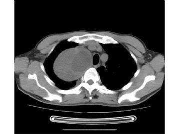
简要病史:  体检CT检查发现右上纵膈占位。主诉无任何症状。实验室常规检查未见异常。
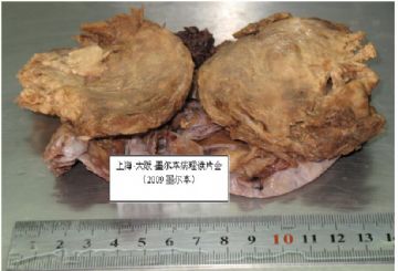
肉眼检查:  巨检请见图片。
  • 右上纵膈肿块(中-日-澳2009墨尔本病理读片会病例1)图1
    图1
  • 右上纵膈肿块(中-日-澳2009墨尔本病理读片会病例1)图2
    图2
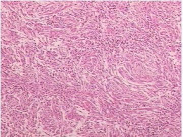
  • 右上纵膈肿块(中-日-澳2009墨尔本病理读片会病例1)图3
    图3
  • 右上纵膈肿块(中-日-澳2009墨尔本病理读片会病例1)图4
    图4
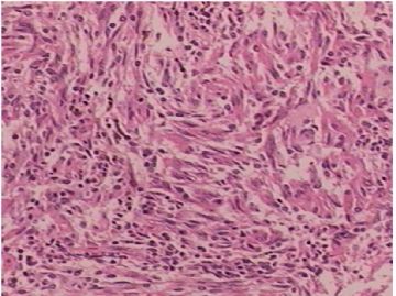
  • 右上纵膈肿块(中-日-澳2009墨尔本病理读片会病例1)图5
    图5
  • 右上纵膈肿块(中-日-澳2009墨尔本病理读片会病例1)图6
    图6
标签:
0
signature
王军臣
×参考诊断
异位脑膜瘤

海上明月 离线

帖子:9476
粉蓝豆:1172
经验:10007
注册时间:2009-08-29
加关注  |  发消息
21 楼    发表于2010-01-20 17:41:00举报|引用
返回顶部 | 快捷回复
本帖最后由 于 2010-01-20 17:42:00 编辑

 以下是陈博士所说的那篇文章的摘要.

Am J Surg Pathol. 2005 Jul;29(7):845-58.

Soft tissue perineurioma: clinicopathologic analysis of 81 cases including those with atypical histologic features.

Hornick JL, Fletcher CD.

Department of Pathology, Brigham and Women's Hospital, and Harvard Medical School, Boston, MA 02115, USA.

Perineuriomas are uncommon benign peripheral nerve sheath tumors that include soft tissue, sclerosing, and intraneural variants. Fewer than 50 soft tissue perineuriomas have been reported to date, and the clinical significance of atypical histologic features is unknown. To characterize these tumors further, 81 soft tissue perineuriomas received between 1994 and 2003 were retrieved from the authors' consult files. Hematoxylin and eosin sections were reexamined, immunohistochemistry was performed, and clinical details were obtained from referring physicians. Forty-three patients were female and 38 male (mean age, 46 years; range, 10-79 years). Tumor size ranged from 0.3 to 20 cm (mean, 4.1 cm) in greatest dimension. Most patients presented with a painless mass. The tumors arose in a wide anatomic distribution: 36 lower limb, 19 upper limb, 15 trunk, 7 head and neck, 3 retroperitoneum, and 1 paratesticular. Forty-two tumors were situated primarily in subcutis, 25 in deep soft tissue, and 9 were limited to the dermis. Nearly all cases were grossly well circumscribed; 12 showed focal microscopically infiltrative margins. Most tumors had a storiform and focally whorled growth pattern; 17 exhibited fascicular areas. Thirty-eight tumors were hypocellular, 15 were markedly hypercellular, and 7 showed alternating zones of hypocellularity and hypercellularity. Stroma was usually collagenous but in 17 tumors was predominantly myxoid, and in 16 was mixed collagenous and myxoid. Mitoses ranged from 0 to 13 per 30 high power fields (mean, 1); 53 tumors had no mitoses. Based on worrisome cytologic or architectural features, 14 cases were classified as atypical perineuriomas: 12 contained scattered pleomorphic cells, 1 showed an abrupt transition from typical morphology to a markedly hypercellular, fascicular area with cytologic atypia, and 1 exhibited diffuse infiltration of skeletal muscle. All tumors were reactive for epithelial membrane antigen; 50 of 78 (64%) expressed CD34, 22 of 76 (29%) claudin-1, 16 of 77 (21%) smooth muscle actin, and 4 of 81 (5%) S-100 protein. All tumors were negative for glial fibrillary acidic protein, neurofilament protein, and desmin. Clinical follow-up was available for 43 patients (mean, 41 months; range, 6-146 months). Among tumors for which the status of surgical margins was known, 52% were widely excised, 31% were marginally excised, and 18% had positive margins. Only two tumors recurred locally (one of which was atypical): one recurred 10 years following primary excision; and one recurred twice, 5 and 9 years following excision. No tumor metastasized. Soft tissue perineuriomas behave in a benign fashion and rarely recur. Atypical histologic features (including scattered pleomorphic cells and infiltrative margins) seem to have no clinical significance and appear to be akin to those seen in ancient schwannoma and atypical (bizarre) neurofibroma.

0
回复
signature
王军臣

海上明月 离线

帖子:9476
粉蓝豆:1172
经验:10007
注册时间:2009-08-29
加关注  |  发消息
22 楼    发表于2010-01-20 09:39:00举报|引用
返回顶部 | 快捷回复
本帖最后由 于 2010-01-20 12:35:00 编辑

 谢谢陈隆文博士对神经束膜瘤的论述,谢谢各位提出的讨论意见。

首先,这个肿瘤是良性的。接着,我们需要进一步鉴别:异位脑膜瘤or神经束膜瘤.

0
回复
signature
王军臣

zsy720121 离线

帖子:33
粉蓝豆:3
经验:38
注册时间:2009-05-03
加关注  |  发消息
23 楼    发表于2010-01-20 08:17:00举报|引用
返回顶部 | 快捷回复
 脑膜瘤 胸腺瘤A型
0
回复

陈隆文博士 离线

帖子:180
粉蓝豆:4
经验:182
注册时间:2008-08-27
加关注  |  发消息
24 楼    发表于2010-01-20 01:42:00举报|引用
返回顶部 | 快捷回复
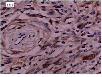
 I guess that with this immunoprofile and the histologic features, one diagnosis in consideration should be Perineurioma. The S-100 protein positive is a little unusual, in Dr. Fletcher's reported 81 soft tissue cases (American Journal Of surgical pathology, 2005, 29:845-858), only 5% were positive for S-100. Most tumors in that paper had a storiform and focally whorled growth pattern (similar to this case).
0
回复

海上明月 离线

帖子:9476
粉蓝豆:1172
经验:10007
注册时间:2009-08-29
加关注  |  发消息
25 楼    发表于2010-01-19 23:47:00举报|引用
返回顶部 | 快捷回复

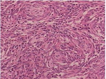
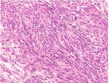
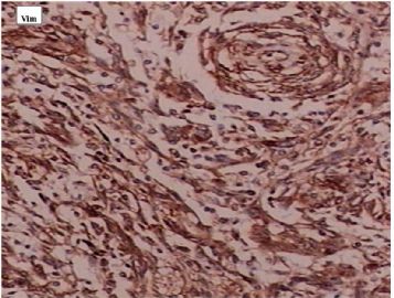
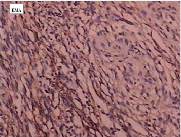
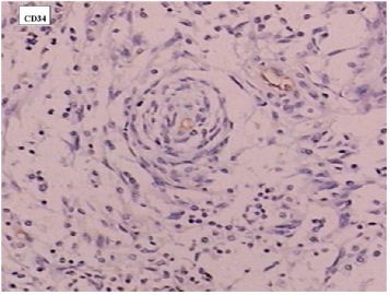
Vim +EMA+S-100+ ;其余均阴性。

  • 图1
  • 图2
  • 图3
  • 图4
0
回复
signature
王军臣

海上明月 离线

帖子:9476
粉蓝豆:1172
经验:10007
注册时间:2009-08-29
加关注  |  发消息
26 楼    发表于2010-01-19 23:19:00举报|引用
返回顶部 | 快捷回复
 Vim +EMA+S-100+CK19-CD21-CD34-CD35-CD68-GFAP -
0
回复
signature
王军臣

wfbjwt 离线

帖子:6628
粉蓝豆:163
经验:7418
注册时间:2006-10-08
加关注  |  发消息
27 楼    发表于2010-01-19 23:16:00举报|引用
返回顶部 | 快捷回复
 S-100和CD1a;CD34和BCL-2;CD68,CD117;EMA,TdT,CKpan,ALK.
0
回复
signature
嫁人就嫁灰太狼,学习要上华夏网。

海上明月 离线

帖子:9476
粉蓝豆:1172
经验:10007
注册时间:2009-08-29
加关注  |  发消息
28 楼    发表于2010-01-19 22:50:00举报|引用
返回顶部 | 快捷回复

  目前大家考虑的有:

(1)低度恶性外周神经鞘膜瘤(恶性神经束膜瘤);(2)SFT;(3)纤维组织细胞瘤;(4)FDC;(5)GIST;(6)异位脑膜瘤,;(7)胸腺瘤;(8)炎性肌纤维母细胞瘤。

那我们再思考一下上述哪些肿瘤中会有图5那样的结构。然后再选择做免疫组化标记就心中有数了。

0
回复
signature
王军臣

wfbjwt 离线

帖子:6628
粉蓝豆:163
经验:7418
注册时间:2006-10-08
加关注  |  发消息
29 楼    发表于2010-01-19 22:29:00举报|引用
返回顶部 | 快捷回复
 首先确定病变性质:低度恶性或良性肿瘤。主要是梭形细胞,混杂炎细胞,形态温和,故考虑滤泡树突细胞肉瘤、炎性肌纤维母细胞瘤。免疫组化有助鉴别。
0
回复
signature
嫁人就嫁灰太狼,学习要上华夏网。

海上明月 离线

帖子:9476
粉蓝豆:1172
经验:10007
注册时间:2009-08-29
加关注  |  发消息
30 楼    发表于2010-01-19 22:19:00举报|引用
返回顶部 | 快捷回复
本帖最后由 于 2010-01-19 22:20:00 编辑
以下是引用michelle_bbmm在2010-1-19 21:43:00的发言:

 图5中有漩涡状的结构


0
回复
signature
王军臣

学浅 离线

帖子:2053
粉蓝豆:79
经验:3125
注册时间:2008-06-01
加关注  |  发消息
31 楼    发表于2010-01-19 22:02:00举报|引用
返回顶部 | 快捷回复
 胸腺瘤AB型
0
回复

96298 离线

帖子:421
粉蓝豆:16
经验:423
注册时间:2009-03-08
加关注  |  发消息
32 楼    发表于2010-01-19 21:57:00举报|引用
返回顶部 | 快捷回复
 FDC,胸腺瘤,异位脑膜瘤,低度恶性外周神经鞘膜瘤
0
回复

michelle_bbmm 离线

帖子:26
粉蓝豆:19
经验:33
注册时间:2009-01-24
加关注  |  发消息
33 楼    发表于2010-01-19 21:43:00举报|引用
返回顶部 | 快捷回复
 图5中有漩涡状的结构
0
回复

ywl 离线

帖子:178
粉蓝豆:12
经验:613
注册时间:2007-02-11
加关注  |  发消息
34 楼    发表于2010-01-19 21:43:00举报|引用
返回顶部 | 快捷回复
 脑膜瘤?
0
回复

michelle_bbmm 离线

帖子:26
粉蓝豆:19
经验:33
注册时间:2009-01-24
加关注  |  发消息
35 楼    发表于2010-01-19 21:41:00举报|引用
返回顶部 | 快捷回复

初学者,前来虚心学习!

希望各位专家老师多多发表意见!

0
回复

fashun 离线

帖子:416
粉蓝豆:26
经验:1257
注册时间:2006-10-10
加关注  |  发消息
36 楼    发表于2010-01-19 17:32:00举报|引用
返回顶部 | 快捷回复
 GIST
0
回复

海上明月 离线

帖子:9476
粉蓝豆:1172
经验:10007
注册时间:2009-08-29
加关注  |  发消息
37 楼    发表于2010-01-19 16:55:00举报|引用
返回顶部 | 快捷回复
 除了梭形细胞交错排列外,请注意图3和图5中有什么不一样的结构。
0
回复
signature
王军臣

dujun0522 离线

帖子:406
粉蓝豆:532
经验:2696
注册时间:2008-07-11
加关注  |  发消息
38 楼    发表于2010-01-19 16:26:00举报|引用
返回顶部 | 快捷回复

 FDC?

0
回复

天天田田 离线

帖子:493
粉蓝豆:5
经验:493
注册时间:2009-05-15
加关注  |  发消息
39 楼    发表于2010-01-19 15:56:00举报|引用
返回顶部 | 快捷回复
本帖最后由 于 2010-01-19 15:58:00 编辑

 1.SFT

 2.纤维组织细胞瘤


0
回复

shandongzhang 离线

帖子:804
粉蓝豆:506
经验:1510
注册时间:2006-09-24
加关注  |  发消息
40 楼    发表于2010-01-19 15:01:00举报|引用
返回顶部 | 快捷回复
本帖最后由 于 2010-01-19 22:43:00 编辑

 先抛砖引玉:

低度恶性外周神经鞘膜瘤(恶性神经束膜瘤)

再仔细看更像FDC

0
回复
回复:42 阅读:7422
共3页/42条首页上一页123下一页尾页
【免责声明】讨论内容仅作学术交流之用,不作为诊疗依据,由此而引起的法律问题作者及本站不承担任何责任。
快速回复
进入高级回复
您最多可输入10000个汉字,按 "Ctrl" + "Enter" 直接发送
搜索回复/乘电梯 ×
按内容
按会员
乘电梯
合作伙伴
友情链接