| 图片: | |
|---|---|
| 名称: | |
| 描述: | |
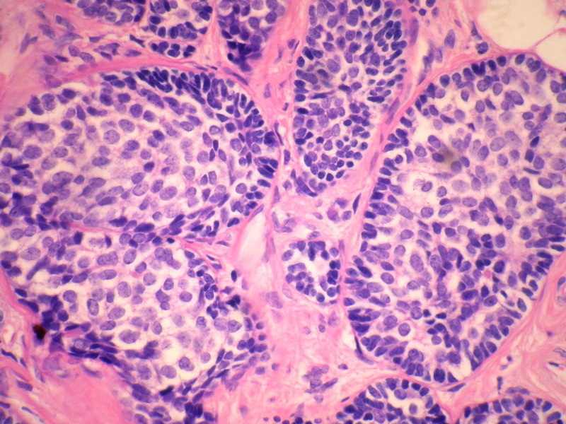
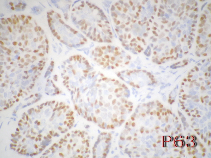
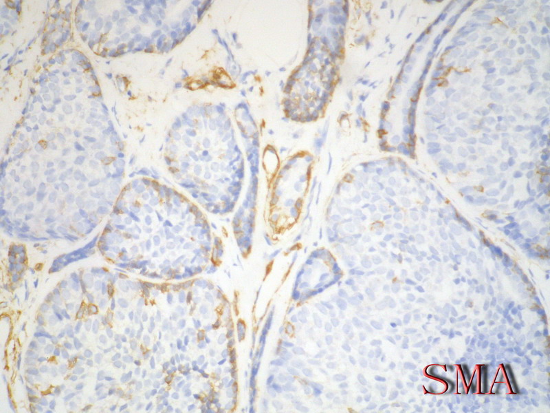
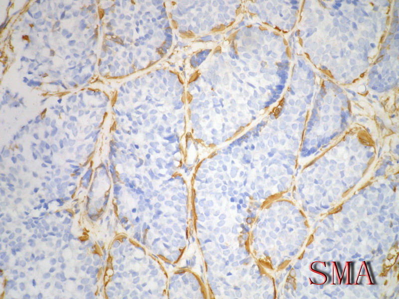
- B228652岁女性,乳腺癌,求类型。已发免疫组化。
| 姓 名: | ××× | 性别: | 年龄: | ||
| 标本名称: | |||||
| 简要病史: | |||||
| 肉眼检查: | |||||
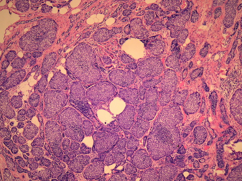
名称:图1
描述:图1
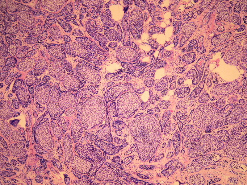
名称:图2
描述:图2
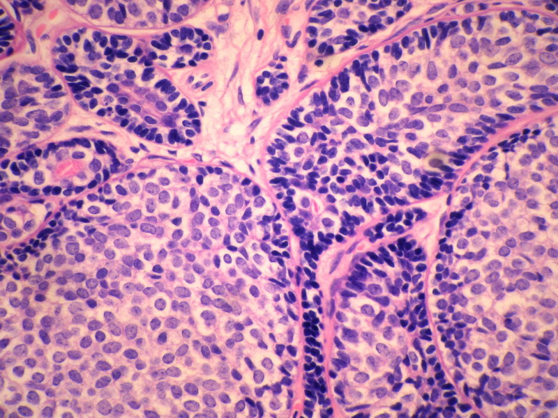
名称:图3
描述:图3
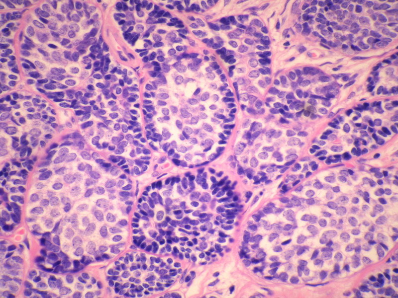
名称:图4
描述:图4
名称:图5
描述:图5
名称:图6
描述:图6
名称:图7
描述:图7
名称:图8
描述:图8
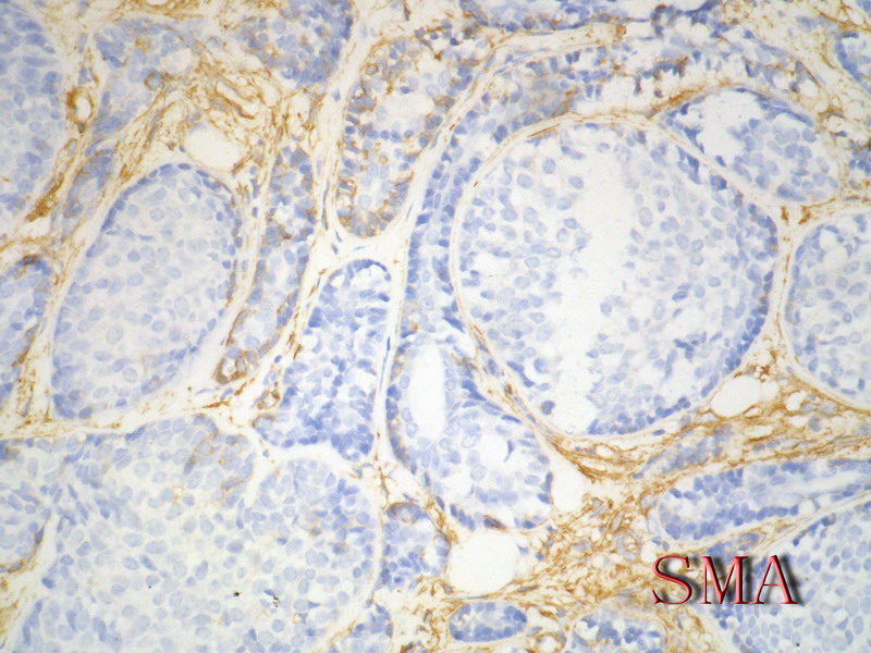
名称:图9
描述:图9
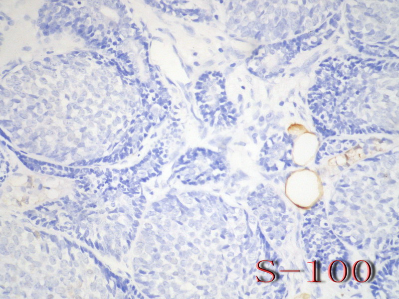
名称:图10
描述:图10
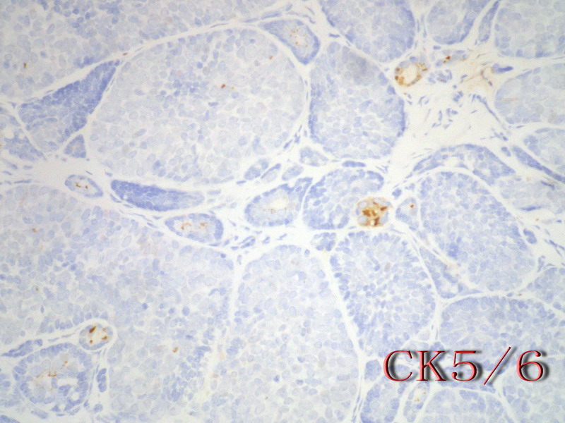
名称:图11
描述:图11
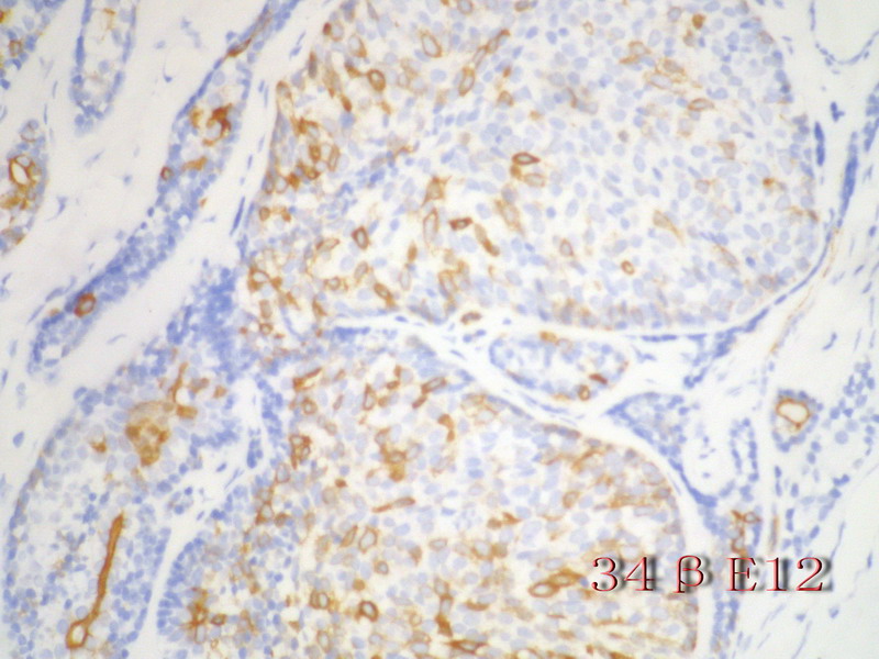
名称:图12
描述:图12
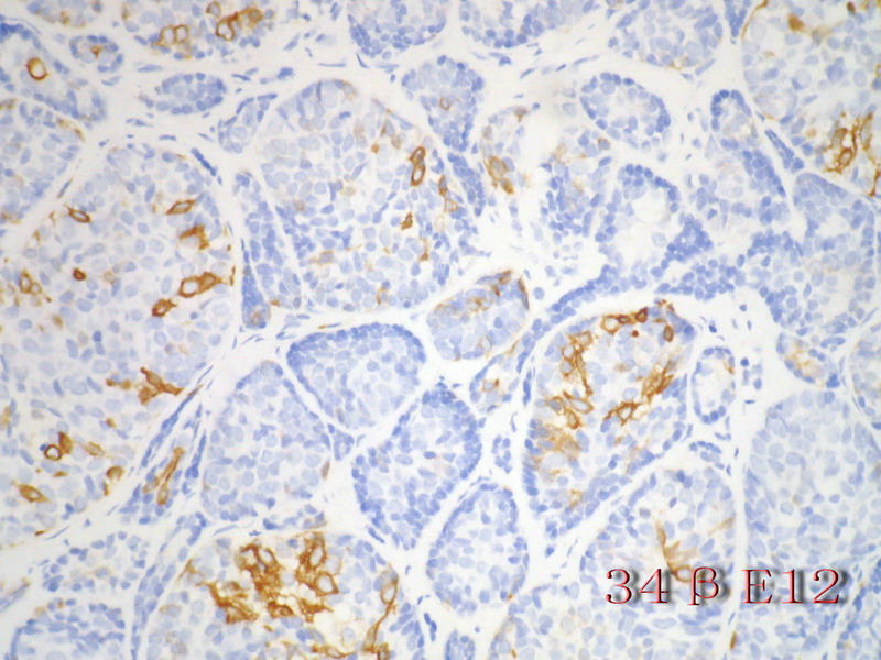
名称:图13
描述:图13
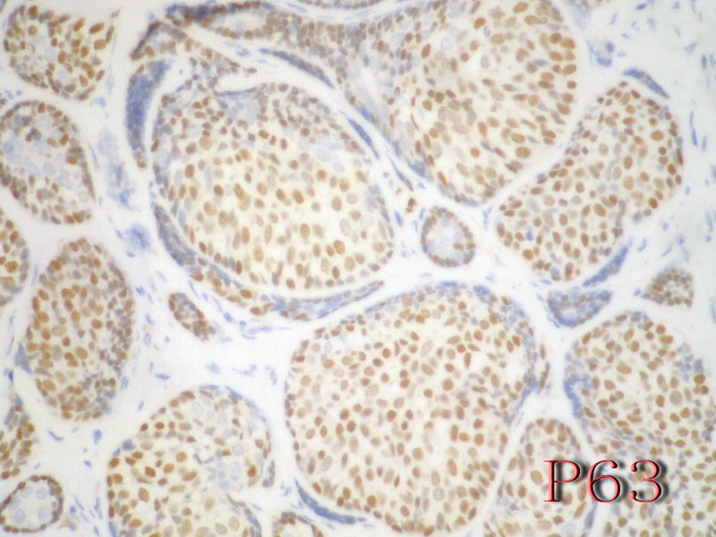
名称:图14
描述:图14
-
本帖最后由 于 2010-09-09 16:26:00 编辑

- 病理,让疾病明明白白。
相关帖子
- • 边界清楚的乳腺包块
- • 右乳肿块,新加免疫组化结果
- • 乳癌类型?
- • 乳腺肿块,请会诊
- • 左侧乳腺外上象限肿物,髓样癌?
- • 乳腺肿块
- • 女,29岁,发现右乳肿块,停止哺乳后手术切除肿块
- • 请求老师会诊,34岁女,乳腺肿物。
- • 乳腺癌类型?
- • 乳腺微创手术活检,诊断?
-
本帖最后由 于 2010-09-09 19:02:00 编辑
Mod Pathol. 2005 Dec;18(12):1623-31.
KIT is highly expressed in adenoid cystic carcinoma of the breast, a basal-like carcinoma associated with a favorable outcome.
Azoulay S, Laé M, Fréneaux P, Merle S, Al Ghuzlan A, Chnecker C, Rosty C, Klijanienko J, Sigal-Zafrani B, Salmon R, Fourquet A, Sastre-Garau X, Vincent-Salomon A.
Department of Pathology, Institut Curie, Paris Cedex, France.
Recent biological studies have classified breast carcinomas into HER2-overexpressing, estrogen receptor-positive/luminal, basal- and normal-like groups. According to this new biological classification, the objectives of our study were to assess the clinical, morphologic and immunophenotypic characteristics of adenoid cystic carcinoma of the breast in order to classify this subtype of breast carcinoma. A total of 18 cases of adenoid cystic carcinoma were identified from the Institut Curie files. Clinical information was available for 16 patients with a median follow-up of 6.5 years. Morphologically, all tumors were graded according to the system defined by Kleer and Oberman (histologic and nuclear grade). Immunophenotype was assessed with anti-ER, PR, HER-2, KIT, basal (CK5/6) and luminal cytokeratins (CK8/18) and p63 antibodies. One out of 18 tumors was nuclear grade 1 (16%), nine were nuclear grade 2 (50%) and eight were nuclear grade 3 (44%). All cases were estrogen receptor, progesterone receptor and HER-2 negative. Epithelial cells were strongly positive around glandular lumina with one or both cytokeratins, identifying the coexistence of CK5/6+ cells, CK5/6 and CK8/18+ cells, CK8/18+ cells and p63+ cells. All cases (100%) were also KIT positive. In all, 15 patients were treated by surgery. Nine of them received adjuvant radiotherapy. Follow-up was available for 16 patients. In all, 14 patients were alive. Two of them, initially treated by surgery only, presented a local recurrence. Two patients died (one of them treated by radiation therapy only died from her disease). Our study shows that adenoid cystic carcinoma of the breast is a special, estrogen receptor, progesterone receptor, HER-2 negative and highly KIT-positive, basal-like breast carcinoma, associated with an excellent prognosis. This highly specific immunophenotype could be useful to differentiate adenoid cystic carcinoma of the breast from other subtypes of breast carcinoma such as cribriform carcinoma.
-
本帖最后由 于 2010-09-09 19:03:00 编辑
Am J Clin Pathol. 2005 Nov;124(5):733-9.
Expression of c-kit in adenoid cystic carcinoma of the breast.
Crisi GM, Marconi SA, Makari-Judson G, Goulart RA.
Department of Pathology, Baystate Medical Center/Tufts University School of Medicine, Springfield, MA 01199, USA.
Breast adenoid cystic carcinoma (BACC) is a biologically distinct tumor with morphologic mimickers, which might make accurate classification problematic. Because c-kit expression has been reported in adenoid cystic carcinoma of various anatomic sites, we evaluated BACC for c-kit by immunohistochemical analysis, comparing the findings to similarly stained mimickers. Tested cases included 6 BACCs, 15 low-grade infiltrating ductal carcinomas (LGIDCs) chosen as potential mimickers, and 15 head-neck adenoid cystic carcinomas (HNACCs). All BACCs showed plasma membranous and cytoplasmic staining equal to or greater than that of adjacent benign epithelium. Five BACCs (83%) expressed c-kit in more than 50% of tumor cells. Only 2 of 15 LGIDCs expressed low-intensity, focal c-kit staining. Of the 15 HNACCs, 10 (67%) expressed c-kit. Hormone receptors were consistently negative in BACCs. All BACCs expressed c-kit, whereas LGIDCs infrequently expressed low-intensity c-kit. Immunohistochemical evaluation for c-kit might aid in accurately classifying carcinomas with histologic features overlapping adenoid cystic carcinoma and LGIDC.
Above abstracts are for some ones who are interested to breast ACC related lesions.
For this case:
It is not like classic ACC. It can be ACC solid type with basaloid features, or BCB.
This kinds of lesions are rare. I just read less than 10 cases of ACC and one or two BCB. I do not have much experience about them. Wish more people share your oppinion.
Also hope 楼主let us know your IHC and interpretation. Thanks, cz
-
本帖最后由 于 2010-01-01 08:59:00 编辑
非常感谢Zhao医师的评论和有关乳腺基底样细胞癌的文献. 我把 "Basaloid carcinoma of breast" 翻译成"乳腺基底细胞样癌" 是为了避免读者将"Basal-like" 与 "Basaloid"混淆., 因为二者有着本质的区别. "Basal-like" 是指免疫表型( CK5/6+和/或CK14+)和基因表达谱与乳腺基底细胞相似, 形态学表现为高级别浸润性导管癌,IHC标记ER-PR-HER2-,临床预后差. 阅读上述文献摘要(未看全文及图片是草率的),作者提出"乳腺基底细胞样癌"是认为乳腺BCB与ACC在形态(无典型ACC)和免疫表型(非肌上皮 SMA-p63-calponin-CD10-)不同,预后比ACC差(3例死于肿瘤,一例带瘤生存).并注意到9例BCB中一例与导管原位癌有过渡;2例伴有3级浸润性导管癌.此外"三阴(triple negative)"的术语应严格用于高级别浸润性乳腺癌免疫表型ER-PR-Her2-的病例,提示预后比"非三阴癌"差. 就此例耒讲:鉴别诊断的焦点主要为:乳腺ACC-实体性, 乳腺BCB,和乳腺恶性圆柱瘤.尚不明确的是Rosen 乳腺病理学第3版第600页Fig 25.21图示中的A-C(Adenpoid cystic carcinoma, high grade: cylindromatous nodules are present in this basoloid tumor) 与 上述文献上"Basoloid carcinoma of breast" 在形态上是否是一回事? 鉴别无疑可用IHC标记,但是对这些少见肿瘤的研究仅是"初级阶段"

- xljin8
| 以下是引用xljin8在2009-11-13 13:14:00的发言: 乳腺腺样囊性(ACC)的形态与唾液腺ACC的形态是相似的。唾腺ACC组织学类型:小管型,筛孔型,实体性,混合型。IHC标记:一般标记CK7+ CK14+ CK17+ CK19+; S-100+ MUC1+ RB+ CD117+: 导管上皮细胞:CEA+EMA+;肌上皮分化细胞:CK+ Vimentin+ MSA+ p63+, SMA+ SMMHC+ Calponin+ GFAP 局限+,等。 ER/PR一般为阴性。 注意:乳腺ACC的形态变异比唾腺ACC更多,而且基底细胞样表型时p63-34BE12+。Ro 等提出乳腺ACC可根据肿瘤内实体成分的百分比分为3级:1级 无实体性成分;2级实体性成分小于30%; 3级实体成分大30%。 本例诊断依据:肿瘤浸润性生长,浸润脂肪组织。主要结构为实体细胞团,但可见小管样结构。实体巢周边细胞栅栏样排列,似汗腺圆柱瘤;二种细胞构成(导管上皮和肌上皮),细胞小,比较均一,无核分裂象,无坏死。鉴别1)浸润性小叶癌无二种类型的细胞,肌上皮应该消失,浸润时呈靶形结构和单行排列。2)浸润性导管癌累及小叶时(小叶癌化)肿瘤细胞异型性大,且应有导管癌成分。 以上个人观点仅供参考。 |

- xljin8


