| 图片: | |
|---|---|
| 名称: | |
| 描述: | |
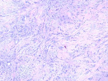
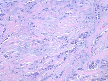
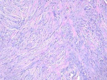
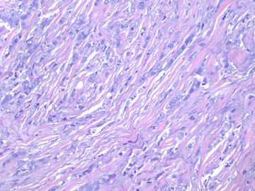
- B2280小叶癌with focal 小管structure(cqz-28)(11-6-09)
| 姓 名: | ××× | 性别: | 年龄: | ||
| 标本名称: | |||||
| 简要病史: | |||||
| 肉眼检查: | |||||
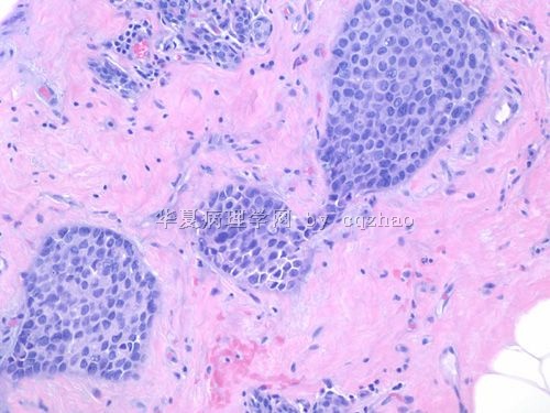
50 岁妇女乳腺肿瘤2.5cm
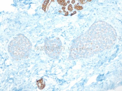
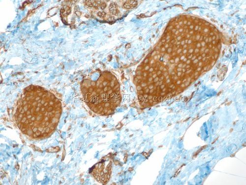
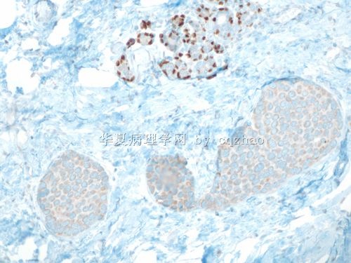
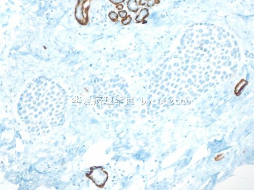
. Fig 1-4. most areas of the tumor.
-
本帖最后由 于 2009-12-16 11:07:00 编辑
相关帖子
- • 女性/45岁 右侧乳腺肿块 诊断?
- • 左乳肿块,新加免疫组化
- • 乳癌?求助
- • 乳腺肿瘤
- • 够小叶癌吗?
- • 女 48岁 左侧乳腺肿块10余天
- • 女性 60岁乳腺肿块 乳腺癌分型
- • 乳腺穿刺
- • 右乳肿块
- • 右乳腺肿块穿刺活检,新加手术后图片
| 以下是引用xljin8在2009-11-18 6:27:00的发言: 2008-04-23 abin医师对浸润性小叶癌做了非常详尽的组织学和细胞学总结, 使我受益非浅。我把它储存在收藏夹量,以便温故知新。 更是敬佩Dr. Chao 乳腺病理诊断的丰富经验和非常感激对国内病理医师的仔细讲解和示教。 |
-
本帖最后由 于 2009-11-18 20:54:00 编辑
2008-04-23 abin医师对浸润性小叶癌做了非常详尽的组织学和细胞学总结, 使我受益非浅。我把它储存在收藏夹量,以便温故知新。
更是敬佩Dr. Chao 乳腺病理诊断的丰富经验和非常感激对国内病理医师的仔细讲解和示教。
对本病例的诊断提出一些问题主要是感到作为一名高年病理医师有责任去帮助求知欲如此高的青年病理医师,去提高他们的诊断思维和拓展他们的知识面。
我个人认为此例就HE形态诊断为混合性浸润性小管小叶癌(Mixed tubulolobular carcinoma)更妥当,是非典型浸润性小叶癌的一种变型(variant)。
请注意 1)典型的小叶癌细胞可以形成微小管-Microtubule(请查阅参考文献中图Fig 9.67)。
2)浸润性小叶癌中可存在小管癌成分(tubular carcinoma), 应诊断为混合性浸润性小管小叶癌(Mixed carcinoma)。
3)混合性癌还包括:浸润性小叶癌 +原位和/或浸润性导管癌( ductal carcinoma)等多种组合。
4)少数浸润性癌在形态学上可具有浸润性导管癌和小叶癌的双重特征或中间特征。因为,形态学表现为浸润性导管癌,而细胞遗传学却具有浸润性小叶癌的分子特点。
参考文献:Diagnostic Surgical Pathology, 5th edition, 2010, p322-323.

- xljin8
| 以下是引用xljin8在2009-11-14 13:17:00的发言:
非常感兴趣的病例,但是我有些疑问: 1)文献中有tubulolobular carcinoma 的名称,应该如何理解? 2)能否提供小管癌区域的免疫组化标记片? 3)诊断时是否因为浸润性小叶癌的恶性程度高而可忽略其他成分? |
Thank for your attention.
1) + 2): will show the IHC results of other areas and have some discussion one by one.
3)诊断时是否因为浸润性小叶癌的恶性程度高而可忽略其他成分?
No. We report all abnormal lesions in the reports in our hospital.
For example:
Invasive ductal or lobular ca
in site ca (DCIS or LCIS)
Atypical lesions: ADH, ALH, atypical papilloma, FEA
Will have one dx line to include all non-neoplastic breast lesions such as FCC, sclerosing adenosis, introductal papilloma, UDH, radial scar, CCC, calcification....
-
本帖最后由 于 2009-11-14 01:00:00 编辑
All of you are right about the tumor.
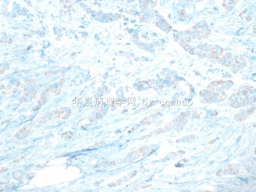
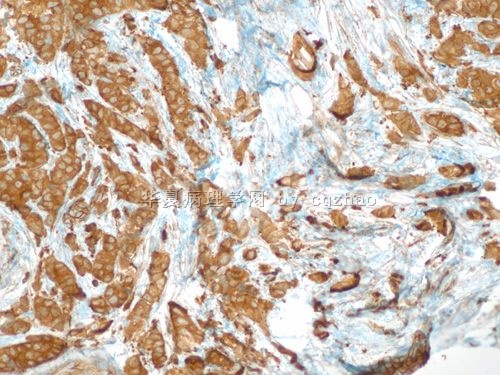
Stains for the main tumor or fig1 (question 1)
E-cad and P120
interpretaion of P120: lobular lesion-cytoplasmic stain; ductal lesion-membrane stain.
We will have the answers for others one by one soon.
名称:图1
描述:图1
名称:图2
描述:图2
-
skyliutong 离线
- 帖子:497
- 粉蓝豆:189
- 经验:878
- 注册时间:2009-02-14
- 加关注 | 发消息
| 以下是引用SOS991229在2009-11-11 23:26:00的发言:
我们在平常工作中也会出现这种情况,我们就冠个总名:浸润性癌,部分区域为浸润性小叶癌,部分区域为浸润性导管癌。不知道赵老师那边是怎样规范的报告?还有是否国外在报告ER,PR时要报%吗?就像ki-67那样+>30%之类的吗?谢谢您! |
We do er/pr/her2/ki67 for all cases of invasive breast ca
I mentioned Her2 report before. I think the Her2 reort is similar among most hospitals in China and the US.
Currently we report ER/PR and ki67 as following.
ER/PR: H score:
example:
ER positive, H score 240 (0 10%; 1+10%; 2+10%; 3+70%)
H score count: (3x70=210)+(2x10=20)+(1x10=10)+(0x10=0) =240
Tumor cell proliferation index (Ki67):
Result 40%,
Index high (low: to 10%; moderate:11-25%; high:26-50%; very high:>50%)
Magee is a breast and gynecologic center. Our reports are very detailed. The report systems can be very variable in different hospitals.
Just for your reference
