| 图片: | |
|---|---|
| 名称: | |
| 描述: | |
- A pap case yesterday-the patient will have biopsy soon
-
本帖最后由 于 2009-09-22 21:56:00 编辑
这是一个非常精彩的病例,尤其是腺细胞的病变,如果从中能学到什么,那将是我们的荣幸。临结尾之时,我提个问题,望不吝赐教。
青青子矜 和掌心0164 在看图二时有分歧(对第二图的分歧在第6和7楼)。
青青子矜 :“图2给我太强烈的羽毛感”
掌心0164 :“但是图2我感觉不像腺,没有那种像羽毛杨的核垂直于细胞团长轴;反而像累腺的鳞状上皮病变。”
最后的结果已经出来了,回过头来看,我们怎么来理解图二呢?顺便谈谈当我们拿起这张液基片时看到这几个细胞的诊断思路及回过头来的总结。
谢谢!

- 朝气用在工作中,热情对待患者去,不屈不挠的韧性用在与学习上,低调做人放在心上。
-
本帖最后由 于 2009-09-23 23:48:00 编辑
| 以下是引用zxh887119在2009-9-22 21:55:00的发言:
这是一个非常精彩的病例,尤其是腺细胞的病变,如果从中能学到什么,那将是我们的荣幸。临结尾之时,我提个问题,望不吝赐教。 青青子矜 和掌心0164 在看图二时有分歧(对第二图的分歧在第6和7楼)。 青青子矜 :“图2给我太强烈的羽毛感” 掌心0164 :“但是图2我感觉不像腺,没有那种像羽毛杨的核垂直于细胞团长轴;反而像累腺的鳞状上皮病变。” 最后的结果已经出来了,回过头来看,我们怎么来理解图二呢?顺便谈谈当我们拿起这张液基片时看到这几个细胞的诊断思路及回过头来的总结。 谢谢! |
青青子矜 and 掌心0164 will give you good answers.
It is very important for cytopathologists to check the histologic follow up results. Review of cytology-histology, histology-cytology, over and over. It is the most important way to learn cytology.
Ok, I need to work on my gyn biopsy for my 54 cases today. Luck part is that i have an excellent gynecologic pathology-fellow to work with me this week.
对照随后的组织学结果对细胞病理医生很重要,不断回顾细胞--组织、组织--细胞,循环往复,这是学习细胞学的最重要方法。
好,我要去处理今天的54例妇科活检了,幸运的是本周我有个能干gynecologic pathology fellow (one year gynecologic fellow after 4 or 5 year pathology resident traning)。
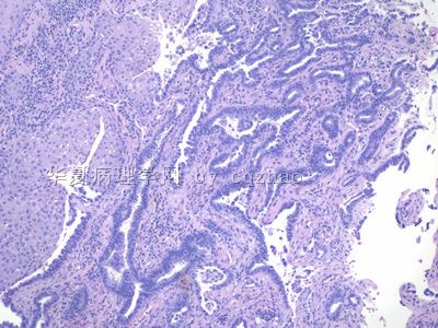
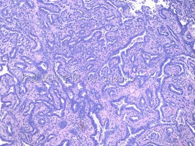
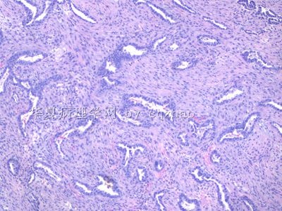
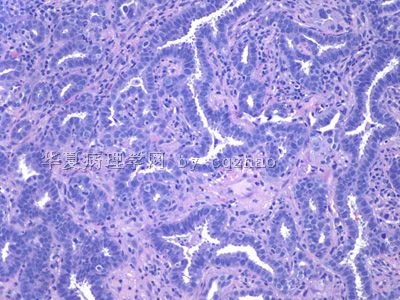
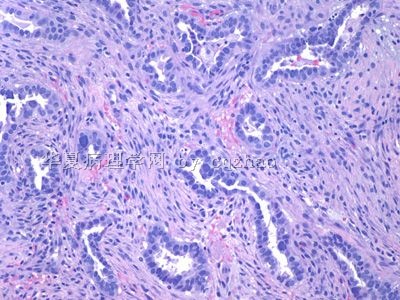
For the histology specimen above we call invasive adenocarcinoma, endometrioid type. The primary can be endocervical or endometrial.
Base on patient's age, Pap smear, and histolgy I personally think it will be an endocervical ca as 青青子矜 mentioned before.
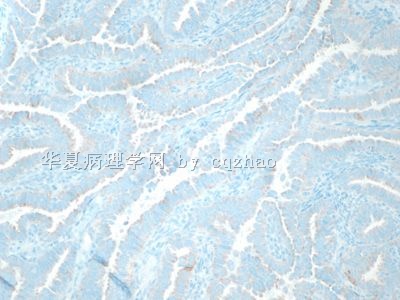
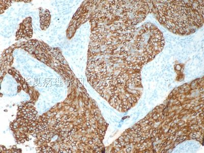
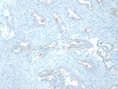
The solid part in the biopsy specimen may be squamous component or adenocarcinoma with solid growth pattern (filling entire glands). I favor it is adenocarcinoma component also. Any way I may order some IHC stains (p63, ck5/6) for education purpose (will not charge the patient).
I will let you know the result when I have it.
-
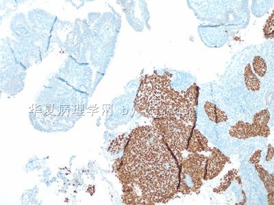
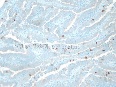
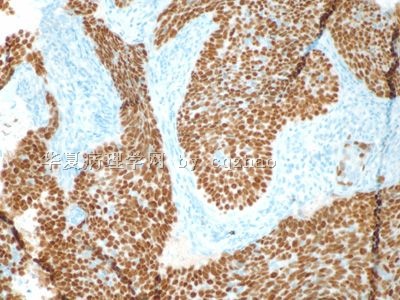
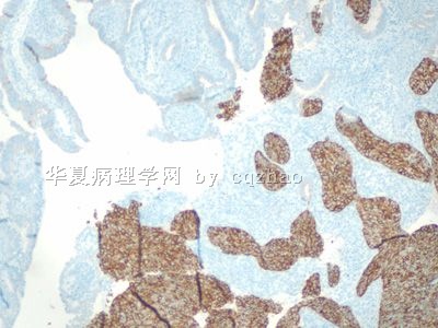
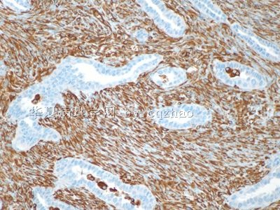
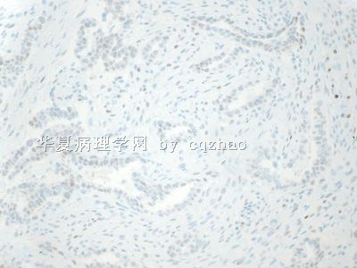
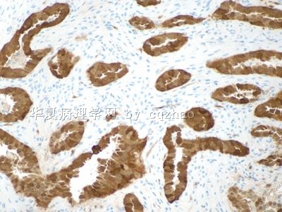
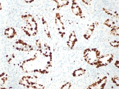
I just got the p63 and ck5/6 stains for the biopsy specimen in floor 28. Are the solid areas poorly differentiated adenocarcinoma with solid growth or squamous cell carcinoma component? Guess what? My original impression is wrong. See above. I will take some photos for your own judgment. I feel this case becomes interesting now.
-
本帖最后由 于 2009-09-24 10:55:00 编辑
回复119老师:
这个病例到目前来看;在组织学中没有看到明显的鳞的病变(前面我说的腺鳞癌;是因为我不懂组织学;对不起大家了)。但是我想等病人全子宫切除之后的组织学结果;因为这里组织学没有宫颈鳞状上皮的组织学结果。目前出来的组织学部位;我们不能完全否定细胞学中是否有鳞状上皮的病变。至少我个人认为到现在为止:第一次赵老师报的AGC-FN和ASC-H是定位准确的。这也是我当时叫的是AGC和ASC-H的原因。我想等所有的结果出来了再好好总结。因为我们在实际工作也遇到过细胞学叫HSIL和AGC的病例。活检:CIN3累腺;做全子宫切除之后还有宫颈管腺癌。虽然这个病例跟我那个病例不一样;但是我目前个人认识水平下,还不能肯定是否合并有鳞的病变。(完全个人观点)

- 掌心0164
Now I think every one should know the diagnosis. 掌心0164 is a 天才. I do not know what you can be if you learn the histology. Ha, ha.
1. Please review the Pap photos again after you know the histologic diagnosis. think over the features of cytology and histology. Learing from the true case is better than learing from the text book.
2. The origin: endocervical or endometrial. Based on all the information above I think we know most likely it is a xxxx tumor. You can fill in the xxxx by yourself.
3. I showed the IHC to the primary pathologist who took case of the biopsy specimen. She wrote an addendum for the case.
This is not a retrospective review case. I am working with your guys together for this case step by step. Sometimes pathology work is interesting.
Thanks, cz


