
图1")
图2")
图3")


| 图片: | |
|---|---|
| 名称: | |
| 描述: | |
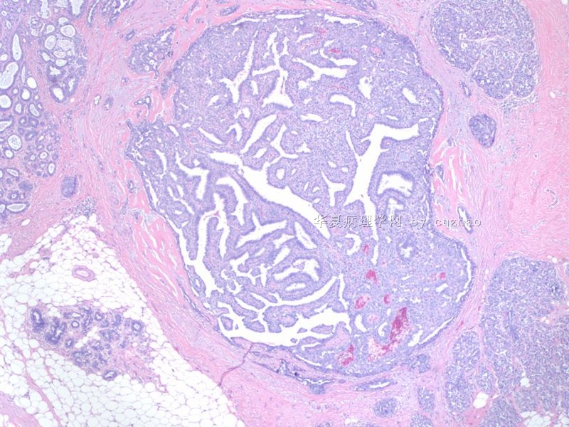
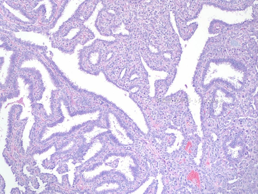
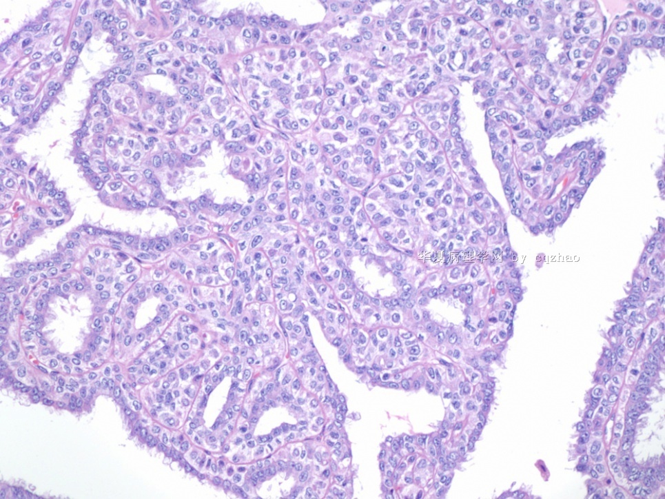
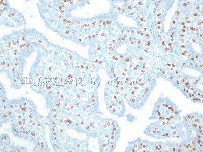
- B2230不典型小叶增生累及乳头状瘤(cqz-27)
同意腺肌上皮瘤的诊断。
乳腺腺肌上皮瘤(adenomyoepithelioma)是一种少见的特殊类型的乳腺良性肿瘤,临床均为女性,发病年龄27~80岁,平均58岁。临床表现主要为乳腺中央或周边部可触及肿块。肿块直径平均1.5~2.5 cm大小,少数可达7 cm。
镜下:肿瘤由增生的肌上皮细胞和腺上皮细胞组成,但与乳腺良性增生病及良性肿瘤中肌上皮细胞增生不同,后者仅围绕导管呈灶性增生,而腺肌上皮瘤则以肌上皮细胞活跃增生为主,细胞弥散,密集呈片,并见轻度异型,增生显著时常压迫腺管,致使管腔闭塞。病变中的肌上皮和腺上皮成分的比例,根据Loose等观察大多为1∶1,少数可达6∶1,且常有一定规律,复发性肿瘤中肌上皮成分明显增多。Tavassoli将此肿瘤分为3型:(1)梭形细胞型:常以肌上皮细胞增生为主,细胞梭形、短梭形呈束状结构。当腺上皮成分少时易与平滑肌瘤混淆。(2)腺管型:本型特点是由腺上皮和肌上皮细胞围绕未稍导管呈聚集性增生,形似一般腺泡、管状或导管腺瘤。当上皮增生显著时,腺管可受压闭塞。此型边缘常不规则,瘤细胞常浸润周围的乳腺组织,因此切除后常易复发。(3)小叶型:增生的肌上皮细胞呈实性、巢状,细胞浆常透明或嗜酸,有的似浆细胞样,它们常围绕受压上皮细胞。肿瘤性肌上皮细胞轻度异型,可见少数核分裂象(1~3/10HPF),瘤周边纤维性包膜向瘤内伸展,把肌上皮分隔呈结节状或小叶状。间质有玻璃样变及钙化。
Accepted Dr. 197's suggestion. I am sending the larger photos. My microscope and camera are new and cost almost US$25,000. However I seldom took good photos. There are too many function I never know.
Humans are more important than eqiupments. Mao once said that victory of a war depends on humans, but not the weapons.
Ok, I need to start to read my today's breast cases now.
| 以下是引用cqzhao在2009-9-4 22:20:00的发言:
Accepted Dr. 197's suggestion. I am sending the larger photos. My microscope and camera are new and cost almost US$25,000. However I seldom took good photos. There are too many function I never know. Humans are more important than eqiupments. Mao once said that victory of a war depends on humans, but not the weapons. Ok, I need to start to read my today's breast cases now. |
谢谢赵老师再次传图!
看来赵老师有一个非常高档的相机,不过,正如赵老师引用的语录所言,人是最重要的因素,所以,我认为上传的好病例伴好照片之精彩的真正缘由是因为上传病例和拍摄照片的是精彩的人!

- “人生没有彩排,每一天都是现场直播”
-
ketty_wang 离线

- 帖子:366
- 粉蓝豆:12
- 经验:520
- 注册时间:2007-09-22
- 加关注 | 发消息
breast lesion (cqz-27) 乳腺病变 (cqz-27)
( )
|
姓 名: |
××× |
性别: |
|
年龄: |
|
|
标本名称: |
| ||||
|
简要病史: |
| ||||
|
肉眼检查: |
| ||||
This is my one today's consult cases
about 60 y/f with breast lesion
What is your diagnosis?
这是我一天的一个会诊病例
大约60岁,女性,乳腺病变
你的诊断是什么?
adenomyoepithelioma. 腺肌上皮瘤
|
Thank above discussion. I especially appreciate the detailed desc ription of 乳腺腺肌上皮瘤by Dr. Zhang. Of cause it does not mean that I agree or disagree the diagnosis. Others can still have your oppinion based on the photos. |
|
感谢以上的讨论。我特别感谢张医生关于乳腺腺肌上皮瘤的详细描述。当然,这并不代表我同意或者不同意这个诊断。其他人可以基于照片持有自己的观点。 |
Accepted Dr. 197's suggestion. I am sending the larger photos. My microscope and camera are new and cost almost US$25,000. However I seldom took good photos. There are too many function I never know.
Humans are more important than eqiupments. Mao once said that victory of a war depends on humans, but not the weapons.
Ok, I need to start to read my today's breast cases now.
我接受了197医生的建议,上传大一些的图片。我的显微镜和照相机都是新的,价值大约25000美元。然而,我很少照到好照片。很多功能我从不知道。人比设备重要的多,毛曾经说过,战争的胜利决定于人,而不是武器。。
好了,我需要开始我今天的乳腺病例读片了。
- 学无止境
-
ketty_wang 离线
- 帖子:366
- 粉蓝豆:12
- 经验:520
- 注册时间:2007-09-22
- 加关注 | 发消息
翻译
breast lesion (cqz-27) 乳腺病变 (cqz-27)
( )
|
姓 名: |
××× |
性别: |
|
年龄: |
|
|
标本名称: |
| ||||
|
简要病史: |
| ||||
|
肉眼检查: |
| ||||
This is my one today's consult cases
about 60 y/f with breast lesion
What is your diagnosis?
这是我一天的一个会诊病例
大约60岁,女性,乳腺病变
你的诊断是什么?
adenomyoepithelioma. 腺肌上皮瘤
|
Thank above discussion. I especially appreciate the detailed desc ription of 乳腺腺肌上皮瘤by Dr. Zhang. Of cause it does not mean that I agree or disagree the diagnosis. Others can still have your oppinion based on the photos. |
|
感谢以上的讨论。我特别感谢张医生关于乳腺腺肌上皮瘤的详细描述。当然,这并不代表我同意或者不同意这个诊断。其他人可以基于照片持有自己的观点。 |
Accepted Dr. 197's suggestion. I am sending the larger photos. My microscope and camera are new and cost almost US$25,000. However I seldom took good photos. There are too many function I never know.
Humans are more important than eqiupments. Mao once said that victory of a war depends on humans, but not the weapons.
Ok, I need to start to read my today's breast cases now.
我接受了197医生的建议,上传大一些的图片。我的显微镜和照相机都是新的,价值大约25000美元。然而,我很少照到好照片。很多功能我从不知道。人比设备重要的多,毛曾经说过,战争的胜利决定于人,而不是武器。。
好了,我需要开始我今天的乳腺病例读片了。
- 学无止境
-
本帖最后由 于 2009-09-05 06:52:00 编辑
Lunch time, Check this caes. Thank Dr. Wang's translation. Now there are a lot of pathologists with high level of English in China. This is a good sign.
似纤毛又非似纤毛: these kinds of structures are very common in columnar cell changes, papilloma, FCC et al. You will notice them often when you pay attention to. Forget these structures. They are not important for the dx of this case.
-
ketty_wang 离线
- 帖子:366
- 粉蓝豆:12
- 经验:520
- 注册时间:2007-09-22
- 加关注 | 发消息
zhao 老师, 您做的工作更多, 我们更应该感谢您才对!
翻译
Lunch time, Check this caes. Thank Dr. Wang's translation. Now there are a lot of pathologists with high level of English in
似纤毛又非似纤毛: these kinds of structures are very common in columnar cell changes, papilloma, FCC et al. You will notice them often when you pay attention to. Forget these structures. They are not important for the dx of this case.
午饭时间,来看看这个病例.感谢王医生的翻译.现在中国国内很多病理学工作者英语水平都很高,这是个好现象.
似纤毛又非似纤毛: 这种结构在柱状细胞变化,乳头状瘤,FCC(?)等病变中都很常见。当你留意的时候,你常常会注意到(这种结构)。忘掉这些结构,这种结构在这个病例的诊断中不重要。
- 学无止境
Interested to ses above differential dx. This is a consult case. The primary pathologist (from a local hospital) thought it maight be a DCIS (papillary type). Some general pathologists in the usa are not good////.
One of our breast path fellow reviewed the case and thought it may be denomyoepithelioma as most of you.


