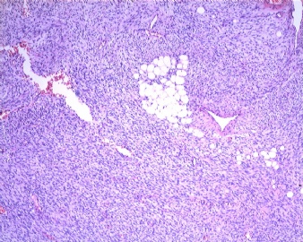
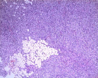
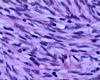
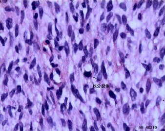
| 图片: | |
|---|---|
| 名称: | |
| 描述: | |
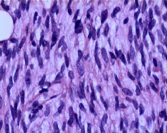
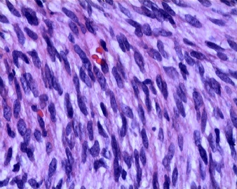
- B1930这例软组织肿瘤各位专家的诊断意见是?
| 姓 名: | ××× | 性别: | 男 | 年龄: | 39 |
| 标本名称: | |||||
| 简要病史: | 右胸壁肿物一年余,大小:3X2CM。 | ||||
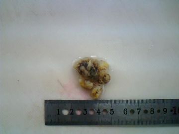
| 肉眼检查: | 灰白灰褐色不整形软组织数块,大小:2X2X1.3CM | ||||
相关帖子
- • 胸壁肿物一例。(隆凸性皮肤纤维肉瘤?)
- • [已确诊]胸壁肿物,请诊断!
- • 胸壁肿瘤
- • 胸壁肿物
- • 左胸壁包块
- • 胸壁肿物
- • 胸壁肿物
- • 【20091218】左胸部皮下肿物
- • 请教:胸壁肿块
- • 左胸壁肿物
-
jianshu322 离线
- 帖子:447
- 粉蓝豆:17
- 经验:1405
- 注册时间:2008-12-22
- 加关注 | 发消息
-
本帖最后由 于 2009-09-26 02:34:00 编辑
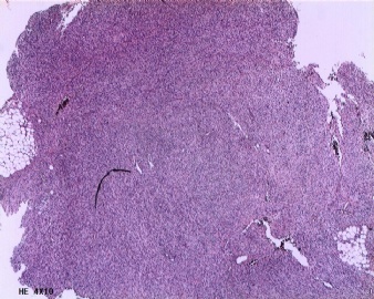
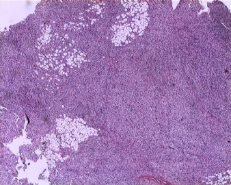
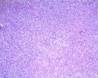
不管是从免疫组化结果还是形态学上,都考虑为低度恶性隆皮纤肉瘤。
对于DFSP的起源有学者认为起源于表达CD34的皮肤树突状细胞或者其亚型,或者DFSP可能是某种特殊类型的表达CD34的神经鞘瘤(May be a peculiar type of nerve sheath tumor since CD34 positive, or may derive from a subset of CD34 positive dermal dendritic cells )。本例CD34(+++),GFAP(+),或许更支持这种说法。
而MPNST(恶性外周神经鞘膜瘤),多发于头颈部及前臂,多数生长于大神经周围体积一般较大可见显著的出血和坏死等,发生于体表皮下者相对少见(至于伴有神经纤维瘤病恶变的病例另论)。本例免疫组化S-100(-)也不支持此诊断。
Bulky deep-seated tumor usually arising from major nerves in neck, forearm, lower leg, buttock。
Gross: large mass producing a fusiform enlargement of a major nerve (often sciatic)Micro: monomorphic serpentine cells, palisading, large gaping vascular spaces, perivascular plump tumor cells, geographic necrosis with tumor palisading at the edges (resembles glioblastoma multiforme),frequent mitotic figures; may have bizarre cells; 15% have metaplastic cartilage, bone, muscle。
Positive stains: S100 (62%), Leu7/CD57 (in neurofibroma-like areas), p53, CD57 (55%), collagen IV, CD99/O13 (86%), protein gene product 9.5,{but PGP9.5 not specific)

- stay hungry,stay foolish.
|
谢谢各位同道给出的意见。以上病理诊断及免疫组化结果是送外院会诊结果:(右胸壁)结合形态学及免疫组化结果:为低度恶性肿瘤,倾向恶性神经鞘膜瘤。 免疫组化:CK(—)、Desmin(—)、SMA小灶(+)、P53(—)、CD34(+)、GFAP(+)。 | ||
|
因各位网友及本人有保留意见,想弄个明白,又拿去另外一家医院会诊,结果:支持纤维肉瘤,低度恶性,结合病变部位,考虑皮肤隆突性纤维肉瘤。请注意切缘及基底部。
|

