| 图片: | |
|---|---|
| 名称: | |
| 描述: | |
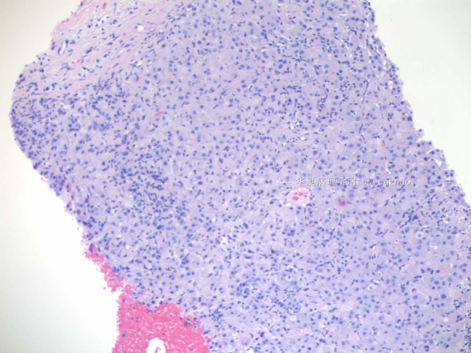
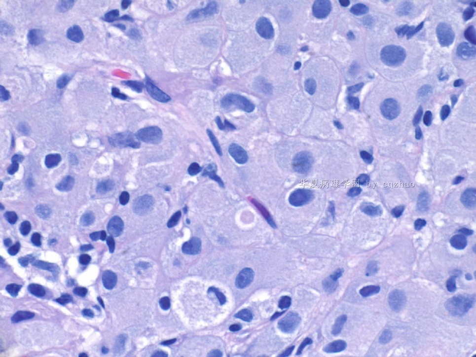
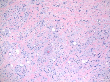
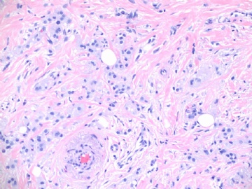
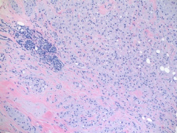
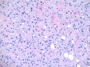
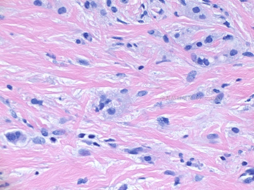
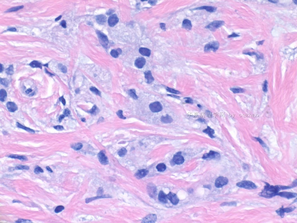
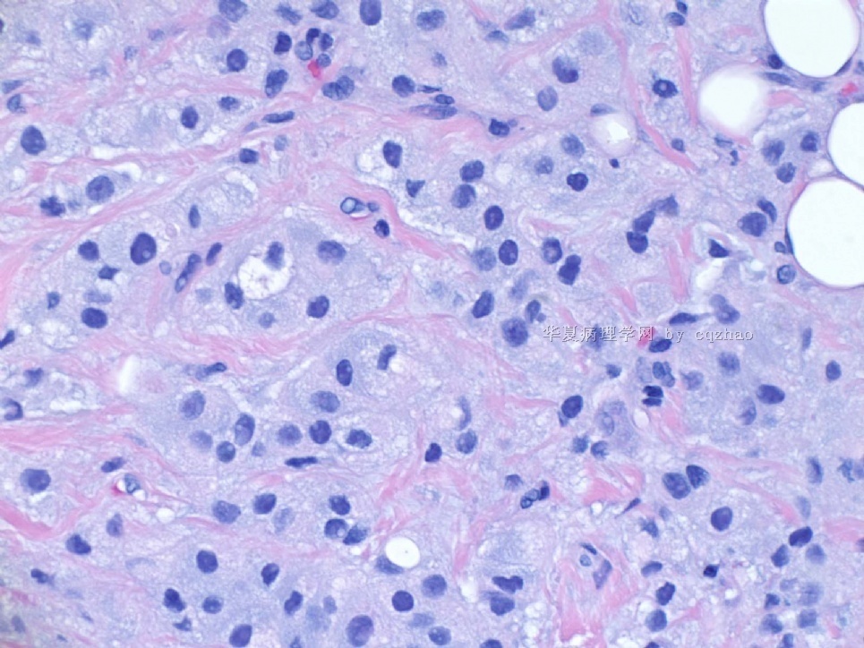
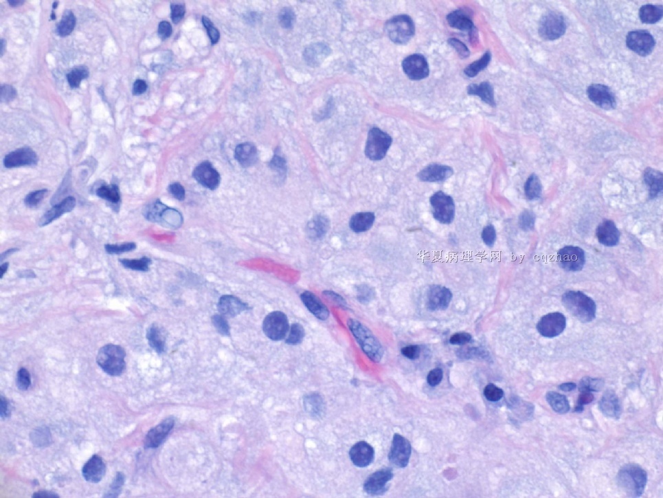
- B2160浸润性导管癌伴大汗腺特征,morphology like histiocytoid or granular cell tumor (cqz-24) 7-16-2009
-
本帖最后由 于 2009-07-21 18:26:00 编辑
http://www.ipathology.cn/forum/forum_display.asp?keyno=147152
Is there any other differential dx
abin译:
请参考http://www.ipathology.cn/forum/forum_display.asp?keyno=147152(组织细胞样型乳腺癌looks like 颗粒细胞瘤 (cqz 17))
是否有其他鉴别诊断?
-
本帖最后由 于 2009-07-21 18:31:00 编辑
tumour cells are diffusely infiltrating in the stroma. There is no obvious glandular structure. However, the cells appear cohesive and frequently clustered together. They have abundant pale blueish (more mucinous-like) cytoplasm instead of eosinophilic granular cytoplasm as in histiocytoid lobular carcinoma. Tumour cells also have ill-defined borders as opposed to lobular carcinoma. So if this is an example of lobular carcinoma, i think it is very atypical. This is a case with an usual appearance. I would consider following differentials: 1. Primary and malligant--- invasive ductal ca. 2. ? Metastatic tumur. 3. ? silicon reaction
abin译:
肿瘤细胞弥漫浸润间质,没有明显腺样结构。然而,细胞有粘附性,多见成簇聚集。胞浆丰富,蓝汪汪(很像粘液样),而不是组织细胞样型小叶癌中的嗜酸性颗粒性胞浆。肿瘤细胞边界不清楚,这与小叶癌也不同。因此,这例如果是小叶癌,我认为是一种非常不典型的病例。本例有少见形态表现,我考虑的鉴别诊断:1、原发性、恶性--浸润性导管癌。2、转移性肿瘤?3、硅胶反应?
反复看图,想寻找蛛丝马迹,Dr.zhao的病例总是那么吸引人啊!谢谢!
此例:在纤维化的背景上,见模糊的粘液样的细胞巢,并浸润在脂肪间,细胞胞界不清,粘液样,核异型性小,未见核分裂,4楼图3略有腺样排列。
1、粘液样的小叶癌?
2、组织细胞样型乳腺癌:胞浆呈细红颗粒状,浆丰富,核异型性很小。CK,E-CA、P120,CD68
3、颗粒细胞瘤:胞浆红颗粒状,内常见神经束。(看到几例,均见到神经束)CD68,S-100,Actin,VIM
4、平滑肌肿瘤粘液样变性:Actin,SMA
5、纤维组织细胞瘤?CD68,VIM
6、转移的粘液细胞肿瘤?PAS染色
7、Pacoma?HMB-45,Malen-A,Actin
8、不会还是血管相关的病变吧?
呵呵,想的太多了,可能超出范围了,不过现在需再了解病史,肿块多大?在乳腺的位置:皮下还是乳腺实质中?患者有无其他部位的病变?病变多长时间了?
此时诊断和鉴别诊断,免疫组化很重要。

- 广州金域病理
-
本帖最后由 于 2009-07-30 14:04:00 编辑
| 以下是引用漫游人在2009-7-21 15:30:00的发言:
tumour cells are diffusely infiltrating in the stroma. There is no obvious glandular structure. However, the cells appear cohesive and frequently clustered together. They have abundant pale blueish (more mucinous-like) cytoplasm instead of eosinophilic granular cytoplasm as in histiocytoid lobular carcinoma. Tumour cells also have ill-defined borders as opposed to lobular carcinoma. So if this is an example of lobular carcinoma, i think it is very atypical. This is a case with an usual appearance. I would consider following differentials: 1. Primary and malligant--- invasive ductal ca. 2. ? Metastatic tumur. 3. ? silicon reaction abin译: 肿瘤细胞弥漫浸润间质,没有明显腺样结构。然而,细胞有粘附性,多见成簇聚集。胞浆丰富,蓝汪汪(很像粘液样),而不是组织细胞样型小叶癌中的嗜酸性颗粒性胞浆。肿瘤细胞边界不清楚,这与小叶癌也不同。因此,这例如果是小叶癌,我认为是一种非常不典型的病例。本例有少见形态表现,我考虑的鉴别诊断:1、原发性、恶性--浸润性导管癌。2、转移性肿瘤?3、硅胶反应? |
-
本帖最后由 于 2009-08-03 20:47:00 编辑
Just complete today's cases. Feel released now. for this weekend night. I will tell you some IHC before I go to home.
Thank all of you for your very reasonable discusion.
AE1/AE3 strongly and diffusely positive.
F1 S-100
F2 CD68.
abin译:
刚完成今天的病例,觉得轻松了。在这周末之夜,在回家前,先告诉大家一些免疫组化结果。
谢谢所有参与讨论的人,你们的观点非常合理。
AE1/AE3 弥漫强阳性
图1 S-100
图2 CD68
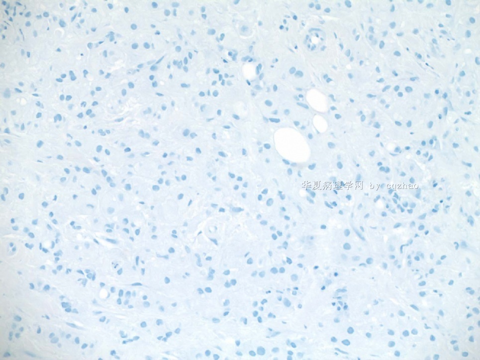
名称:图1
描述:图1
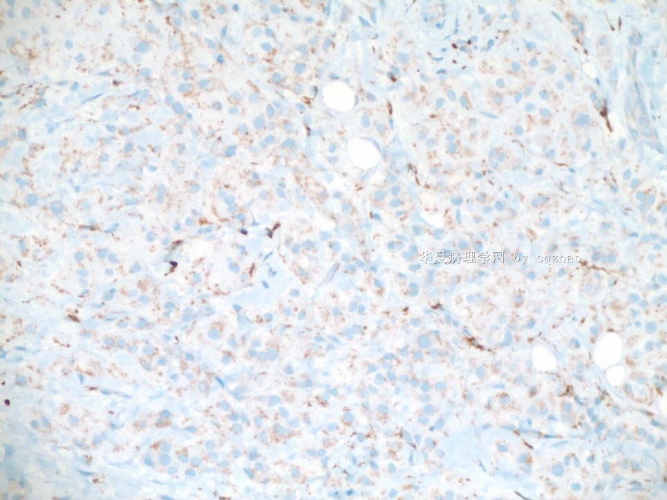
名称:图2
描述:图2



 支持向你学习!


