| 图片: | |
|---|---|
| 名称: | |
| 描述: | |
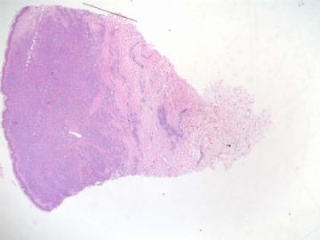
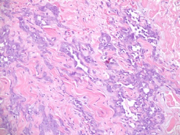
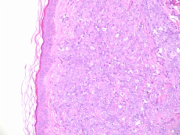
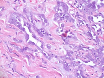
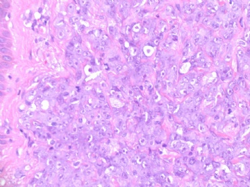
- B1839乳房高级别血管肉瘤 (cqz-21)
Thank abin to send the table above.
We can find a lot of tables from different expert text book. In practice low grade tumor needs to differ from benign or atypical hemagioma. Low grade tumor is easy to distinguish from intermediate or high grade tumor. However it is difficult to tell the difference between the intermediate grade tumor and high grade tumor for some cases. Fortunately it is not clinical important to distinguish intermediate grade tumor from high grade tumor.
-
本帖最后由 于 2009-07-09 12:14:00 编辑
 还是套用这个表:
乳腺血管肉瘤的组织学特征(from Rosen Breast Pathology)
组织学特征 低级别 中级别 高级别
病变累及乳腺实质 有 有 有
吻合的血管腔 有 有 有
深染的内皮细胞 有 有 有
内皮细胞成簇 极少 有 显著
乳头形成 无 局灶 有
实性和梭形细胞灶 无 无或极少 有
核分裂 罕见或无 见于乳头区 多;低级别区域也有
血湖 无 无 有
坏死 无 无 有
--------------------------------------------------------------------------------------------------------------------------
本例:无坏死,无血湖,核分裂多,有实性区和梭形区……归入中-高级别血管肉瘤。
我觉得乳腺的血管病变有部位特殊性,因此我接受乳腺病理专家Rosen的标准。软组织病理专家提出的血管肉瘤的诊断标准可能不完全适用于乳腺(未深入学习过,猜测而已,以后有机会比较一下)。

华夏病理/粉蓝医疗
为基层医院病理科提供全面解决方案,
努力让人人享有便捷准确可靠的病理诊断服务。
Thank Dr. Zhang, thank you for your input.
Dr. Rosen's paper indicated that ki67 may be useful in distinguish low grade angiosarcoma from benign hemagioma. Last week I asked the question to Dr. Christopher Fletcher, Professor and Director of Surgical Pathology, Brigham & Women's Hospital, Harvard medical School. He answered that We do not use Ki-67 in this context.
Anyway ki67 stain is not useful for dx of high grade or intermediate grade angiosarcoma.
-
本帖最后由 于 2009-07-07 09:53:00 编辑
High grade angiosarcoma. See the discussion in cqz-case 22. I do not think pathologists need to separate angiosarcomas arising from breast skin or from breast parenchyma. Generally in postradiation angiosarcomas, cutaneous presentation are more common.
| 以下是引用cqzhao在2009-7-7 9:59:00的发言: Just noted that Dr. zhang mentioned above Ki67 for differentiation cutanuous angiosarcoma from paranchymal angiosarcoma. Could you show the reference? Thanks, cz |
