| 图片: | |
|---|---|
| 名称: | |
| 描述: | |
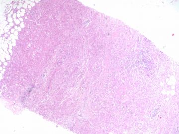
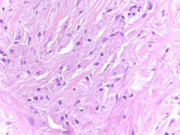
- B1786组织细胞样型乳腺癌looks like 颗粒细胞瘤 (cqz 17)
-
Virchows Arch. 2007 Apr;450(4):397-403. Epub 2007 Feb 28.
-
Expression of aberrant mucins in lobular carcinoma with histiocytoid feature of the breast.
Pathology Section, Kanazawa University Hospital, 13-1 Takara-machi, Kanazawa, Ishikawa, 920-8641, Japan. yzen@med.kanazawa-u.ac.jp
The clinicopathological profiles of histiocytoid carcinoma of the breast have not been well examined because of their rarity and heterogenous groups of ductal and lobular origin. A large foamy or granular cytoplasm of histiocytoid carcinoma was characterized by abundant mucin, but the properties of mucin in histiocytoid carcinoma have also not been well investigated. We selected eight cases of histiocytoid features of invasive lobular carcinoma (HLC) and compared with 14 age- and tumor size-matched cases of classical invasive lobular carcinoma (CLC). Mucin profiles were significantly different between the two groups: a fair number of HLC cases were immunopositive for MUC2 and MUC5AC (75 and 50%, respectively); in contrast, almost all CLC cases showed both as negative. Both groups were immunopositive for MUC1 and negative for MUC4 and MUC6. The prognosis of HLC was significantly worse than CLC; HLC showed shorter disease-free time than CLC (p=0.0262). In particular, HLC with MUC2 and MUC5AC expressions showed significantly shorter disease-free time and survival time than lobular carcinoma without the expressions of MUC2 and MUC5AC (p=0.0055 and p=0.0060, respectively). Therefore, the expression of 'non-mammary mucins', such as MUC2 and MUC5AC in HLC, is characteristic and indicates the more malignant transformation of tumor cells and poorer prognosis.
| 以下是引用cici在2009-5-1 20:04:00的发言: 多谢 cqzhao老师提供的好病例,我们总是担心颗粒细胞瘤过诊断为乳腺癌,也应该想到会有组织细胞样型癌低诊断成颗粒细胞瘤的可能,两种误诊都会产生严重后果,最谨慎的方法是不要100%相信组织形态,对于不常见的组织类型一定要做免疫组化证实后再发报告。 |
You are exactly right. As pathologists We must feel we are sure and believe it is 100% right when we sign out the cases with our diagnosis,especially for some malignant cases.
We need to use all available methods to rule in or rule out our differential dx when we are not 100% sure.We can use some acessary methods (like IHC), showing the cases to your coleques, sending out to experts for consulation et al. Of cause we know that no persons are always 100% right. Also we know the limitation of the pathology. If we use all your possible sources and if we still do not know the dx, we can have the differential dx in the pathology report with a long comment.
Anyway we cannot guess dx, cannot think 99% of chance will be xxx for your dx. This is the priciple we all pathologists should observe.
组织细胞样乳腺癌,瘤细胞胞浆丰富,红色颗粒状或泡沫状,核小,圆形,偏位,与颗粒细胞瘤确实不易鉴别。IHC很重要。
还想请教赵博士如下问题:
在HE上
1、组织细胞样乳腺癌与颗粒细胞瘤:间质反应有差别吗?(一个良性,一个恶性)
2、组织细胞样乳腺癌与颗粒细胞瘤:瘤细胞巢排列有无差异?
3、组织细胞样乳腺癌与颗粒细胞瘤:肿瘤边界如何?生长方式如何?
4、均与黄色瘤怎样鉴别?与吞噬脂质的细胞肿瘤怎样鉴别?
5、与退化的平滑肌瘤怎样鉴别?(我们见到一例食管的颗粒细胞瘤与平滑肌瘤相似,IHC确诊的),乳腺平滑肌瘤少见,提此问题仅想了解形态上的差异)。
呵呵,因为此类病例看的少,提的问题多了,请赐教!再次感谢!

- 广州金域病理
| 以下是引用天山望月在2009-5-2 17:44:00的发言:   组织细胞样乳腺癌,瘤细胞胞浆丰富,红色颗粒状或泡沫状,核小,圆形,偏位,与颗粒细胞瘤确实不易鉴别。IHC很重要。 还想请教赵博士如下问题: 在HE上 1、组织细胞样乳腺癌与颗粒细胞瘤:间质反应有差别吗?(一个良性,一个恶性) 2、组织细胞样乳腺癌与颗粒细胞瘤:瘤细胞巢排列有无差异? 3、组织细胞样乳腺癌与颗粒细胞瘤:肿瘤边界如何?生长方式如何? 4、均与黄色瘤怎样鉴别?与吞噬脂质的细胞肿瘤怎样鉴别? 5、与退化的平滑肌瘤怎样鉴别?(我们见到一例食管的颗粒细胞瘤与平滑肌瘤相似,IHC确诊的),乳腺平滑肌瘤少见,提此问题仅想了解形态上的差异)。 呵呵,因为此类病例看的少,提的问题多了,请赐教!再次感谢! I wish you can find the book and answer the questions and share with us. As you mentioned, you must do the IHC for diagnosis of these lesions.  |


N69%0VAWBANRVH8U(E.jpg)


