| 图片: | |
|---|---|
| 名称: | |
| 描述: | |
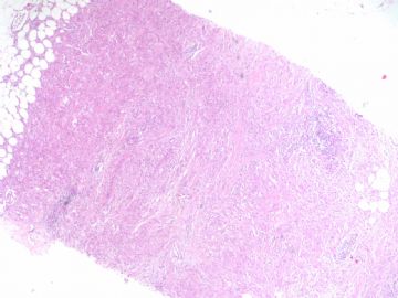
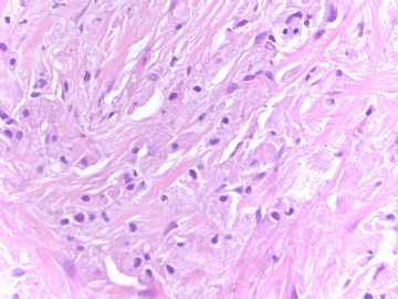
- B1786组织细胞样型乳腺癌looks like 颗粒细胞瘤 (cqz 17)
| 以下是引用笃行者在2009-4-27 17:29:00的发言: 该例首先考虑颗粒细胞瘤,但是在做诊断之前必须进行S-100蛋白和CK标记,以排除组织细胞样型乳腺癌(或称肌母细胞样癌) |
Above should be the answer from pathologists.
You are wrong if you called GCT directly even though this case were a GCT. I mentioned many times in this web that we are pathologists and we cannot guess dx.
-
本帖最后由 于 2009-04-30 03:51:00 编辑
In fact this is a very easy and unusual case which I had when I was at AFIP. I first saw the case. Clearly it was granular cell tumor by cytomorphology. Tumor cells show small round and nuiform nuclei and abundent and granular eosinophilic cytolasm, classic features of GCT. Surprisingly IHC stains indicated the tumor cells were strongly positive for CK7, AE1/AE3, and negative for S-100, CD68. So it is histiocytoid carcinoma, a variant of lobular carcinoma.
Of cause I am sure all of us know that these two tumors have much different prognosis.
The lession for this case is that pathologists must rule out all other possible differential diagoses before we make our final diagnosis in our clinical practice. This priciple should be used for all cases even though we think we know the dx of the cases.
-
本帖最后由 于 2009-06-03 21:03:00 编辑
Ann Diagn Pathol. 2002 Jun;6(3):141-7.
-
E-cadherin immunohistochemical analysis of histiocytoid carcinoma of the breast.
Department of Pathology and Laboratory Medicine, The University of Texas Health Science Center, Houston, TX, USA.
Histiocytoid carcinoma is a rare type of invasive breast carcinoma. It has been considered to be a variant of lobular carcinoma, a variant of apocrine ductal carcinoma, and an apocrine variant of lobular carcinoma and to resemble lipid-rich carcinoma. In attempts to elucidate its histogenesis, investigators have used mucin and oil red O histochemical analysis and GCDFP-15 immunostaining. E-cadherin is a relatively recent addition to the armamentarium of immunohistochemical markers used for cell differentiation and is a member of a family of transmembrane glycoproteins that has been shown to have a strong correlation with the histologic phenotypes of breast carcinoma. Most ductal carcinomas show diffuse membrane expression of E-cadherin, and lobular carcinomas are characterized by complete lack of membrane staining of E-cadherin. The object of this study was to use E-cadherin immunohistochemical analysis to help clarify the histogenesis of histiocytoid carcinoma. Fourteen cases containing the diagnosis of histiocytoid carcinoma of the breast were identified at M. D. Anderson Cancer Center (Houston, TX) from 1988 to 2001. All cases were rereviewed, histologic features were evaluated, and immunohistochemical staining with E-cadherin and GCDFP-15 was performed. Clinical information was extracted from the patients' medical records. Eleven cases met published histologic criteria for histiocytoid carcinoma. The remaining three cases were apocrine carcinoma. The pattern of tumor infiltration was solid, without secondary lumen formation in all cases of histiocytoid carcinoma. Lobular carcinoma in situ was identified in eight cases, but was absent in three. There was no E-cadherin immunohistochemical staining in eight of the 11 cases of histiocytoid carcinoma (72.7%). GCDFP-15 was immunoreactive in all 10 cases of histiocytoid carcinoma where it was performed. Follow-up data was available for nine of the 11 cases of histiocytoid carcinoma: six patients were alive with disease at 1.5 to 48 months, one patient had died of disease at 60 months, and two patients had no evidence of disease at 32 and 45 months. We conclude that histiocytoid carcinoma has an immunophenotypical profile consistent with both ductal and lobular differentiation. Moreover, the lack of consistent morphologic features, a specific clinical profile, and a distinct immunohistochemical pattern lead us to hypothesize that histiocytoid carcinoma is not a special type of breast cancer. Copyright 2002, Elsevier Science (USA). All rights reserved.
组织细胞样乳腺癌E-钙粘素免疫组织化学分析
组织细胞样乳腺癌是一种少见类型的浸润型乳腺癌。有人认为它是小叶癌的变型、有顶浆分泌特征导管癌(大汗腺癌?)的变型和有顶浆分泌特征小叶癌的变型,还有人认为它类似于富于脂质癌。研究者曾做粘液素和油红O组织化学及GCDFP-15 免疫组织化学分析,尝试阐明它的组织来源。E钙粘素是最近增加的一种用于确定细胞分化的免疫组织化学标记,它是跨膜糖蛋白家族的一员,并且被证实与乳腺癌的组织学表型有强相关性。大多数导管癌E钙粘素表现为散在的膜阳性,而小叶癌E钙粘素标记则完全缺乏这种膜阳性。本研究的目的是用E钙粘素免疫组织化学分析来帮助阐释组织细胞样癌的来源。从1998年至2001年,有14例在Anderson癌症中心被证实诊断为组织细胞样乳腺癌的病例,所有病例都被重新复习,重新评价组织学特征,均做E钙粘素和GCDFP-15 免疫组织化学标记,临床信息摘自病人的病案记录,11例符合组织细胞样乳腺癌的组织学诊断标准,其他三例为大汗腺样癌。在所有的组织细胞样癌的病例中,肿瘤浸润模式为实性,没腔隙结构。有8例伴有小叶原位癌,其他三例没有。11例组织细胞样癌中9例有有效的随访资料,6例带瘤生存1.5到48个月,1例在第60个月死亡。我们得出结论组织细胞样癌既导管癌的免疫组织学特征又有小叶癌的免疫组织学特征。而且由于缺乏一致的形态学特点和缺乏特有性的临床表现以及特征性的免疫组织化学表现,我们猜测组织细胞样乳腺癌不是乳腺癌的一种特殊类型。
-
Ann Pathol. 2003 Jun;23(3):249-52.
-
Histiocytoid breast carcinoma: a case report of an uncommon histologic variant of lobular carcinoma.
Département de Pathologie, Faculté de Médecine, Université Aristote de Thessalonique, 54006 Thessalonique, Grèce.
A rare case of invasive histiocytoid breast carcinoma is presented. A middle aged female patient underwent quadrantectomy for a palpable mass in her right breast. The neoplastic cells showed pronounced histiocytoid appearance and immunopositivity for cytokeratins 7 and CAM 5,2, EMA, CEA, GCDFP-15, MFG and ER/PR. We report here this uncommon histologic pattern because of the considerable diagnostic interest. Recognition of this histologic variant either in primary or metastatic locations may present some difficulties to the pathologist. Differential diagnostic problems are emphasized and the literature is reviewed.
-
Ann Pathol. 2004 Jun;24(3):259-63; quiz 227.
-
[Infiltrating lobular carcinoma of the breast with histiocytoid features: three cases.]
[Article in French]Laboratoire d'Anatomie et Cytologie Pathologiques Marcel Mérieux, Avenue Tony Garnier, 69007 Lyon. maugros@lab-merieux.fr
Histiocytoid carcinoma of the breast, a cellular variant of invasive breast cancer, is mainly found among infiltrating lobular carcinomas (ILC). It can be easily confused with benign conditions or other mammary tumors also composed of cells with a pink granular to foamy cytoplasm and an eccentric nucleus. We report 3 cases of histiocytoid ILC. Our aim is to discuss recent immunocytochemical data that could suggest a special type of apocrine differentiation of tumor cells, including a diffuse immunoreactivity for GCDFP-15 (Gross Cystic Disease Fluid Protein 15) and a predominant expression of androgen receptor, and to describe the features useful for the differential diagnosis.
-
Pathol Int. 2005 Jun;55(6):353-9.
-
Histiocytoid breast carcinoma: solid variant of invasive lobular carcinoma with decreased expression of both E-cadherin and CD44 epithelial variant.
Department of Pathology, Kensei General Hospital, Iwase, Nishi-Ibaraki, Japan.
Histiocytoid breast carcinoma (HBC) is a rare type of breast carcinoma with morphologic characteristics resembling those of histiocytes. Described herein are cytological and histological findings in a case of HBC. Fine-needle aspiration cytology revealed numerous loosely cohesive tumor cells with abundant foamy to granular cytoplasm and bland-appearing nuclei. The resected tumor exhibited a solid growth pattern instead of classic invasive lobular patterns observed in most reported cases of HBC. However, distinct intracytoplasmic lumina and Pagetoid extension to ducts suggested that this tumor was a variant of invasive lobular carcinoma. To determine the cause of the loose cellular cohesiveness of this HBC, its expression of the epithelium-related cell adhesion molecules E-cadherin and CD44v8-10 (CD44 epithelial variant) was examined. Immunohistochemically, E-cadherin was not detected, similar to most lobular carcinomas. Furthermore, competitive reverse transcription-polymerase chain reaction (RT-PCR) analyses among alternatively spliced variants of CD44 revealed that the ratio of expression of CD44v8-10 to that of CD44v10 (dominant variant in leukocytes) was lower than that for the reference breast carcinoma samples. It is concluded that the present case of HBC was a solid variant of invasive lobular carcinoma exhibiting foamy to granular cytoplasmic change. Decreased expression of both E-cadherin and CD44 epithelial variant may be responsible for the loose cellular cohesiveness observed in HBC.
-
J BUON. 2006 Jul-Sep;11(3):359-62.
-
Histiocytoid breast cancer: an uncommon histologic variant of lobular carcinoma.
Department of General Surgery and Surgical Oncology, Numune State Hospital, Konya, Turkey. aydaneroglu@hotmail.com
We present a case of histiocytoid breast cancer (HBC), which is a rare variant of breast carcinoma. The patient underwent an excisional biopsy for a palpable mass in her right breast. The neoplastic cells exhibited pronounced histiocytoid appearance. Immunohistochemical and histochemical studies were performed and tumor cells were positive for PAS, keratin PAN-CK, EMA, progesterone (PgR) and estrogen receptors (ER), while S-100 protein, CD68, CEA, and c-erbB2 were negative. Differential diagnosis issues and immunohistochemical data are emphasized and the literature is reviewed.
-
lfl001200546 离线
- 帖子:2808
- 粉蓝豆:40
- 经验:2808
- 注册时间:2007-02-14
- 加关注 | 发消息


