| 图片: | |
|---|---|
| 名称: | |
| 描述: | |
- B1581Breast lesions cqz (12) (2-16-2009)
| 姓 名: | ××× | 性别: | 年龄: | ||
| 标本名称: | |||||
| 简要病史: | |||||
| 肉眼检查: | |||||
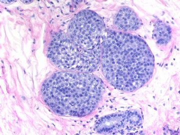
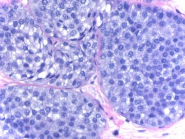
Breast core biopsy with 8 cores. There are multiple breast lesions. Try you diagnosis one by one.
Lesion 1 20x and 400x
-
本帖最后由 于 2009-03-19 23:29:00 编辑
相关帖子
- • 乳腺癌?
- • 女性 冰冻为乳腺浸润性导管癌,现切除标本,肿块旁组织
- • 女性 33岁 乳腺肿块
- • 乳腺包块
- • 乳腺两个相邻导管内的病变
- • 乳腺肿物
- • 38岁乳腺(新加HE切片)
- • 乳腺包块。33岁
- • 左乳肿块,协助诊断
- • 乳腺肿物求助
-
alading1999 离线
- 帖子:63
- 粉蓝豆:31
- 经验:562
- 注册时间:2009-01-17
- 加关注 | 发消息
Seem most of you agree the interpretation of F 1-4, 6. You can give your reasons if you do not agree.
F 5, 7involve columinar cell change, 平坦型上皮非典型性(FEA) and related lesions. FEA is a very controversial term. There are many issues which are not clear, such as dx criteria, managment: present in in core biopsy, in excisional bx, .in surgical margins. In fact even in the US, many general surgical pathologists do not know the term or do not know how to make the diagnois. Of cause most of surgens know nothing about FEA. We have alot of consult cases from other hospitals. Often we need to talk with primary pathologists. This is why I know a lot of general pathologists do not know the term well.
Interesting to know that our pathologists in China use this term or not. Please share your knowledge about FEA with ours.
Thanks
Yes, We use the terminology of columnar cell lesion in diagnosis.
The columnar cell lesion especially flat cell atypia is related to some low grade carcinoma such as tubular carcinoma, tubulobular carcinoma etc. However, I am puzzled about FEA accompany with structure complex, should it be diagnosed as atypic duct hyperplasia or FEA, just as figures shown in lesion 7? According to the definition of FEA there should not have structure complex.
Interested in the topic very much.
Thanks!


