| 图片: | |
|---|---|
| 名称: | |
| 描述: | |
- B1581Breast lesions cqz (12) (2-16-2009)
| 姓 名: | ××× | 性别: | 年龄: | ||
| 标本名称: | |||||
| 简要病史: | |||||
| 肉眼检查: | |||||
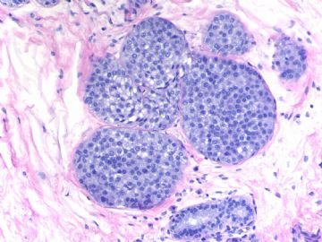
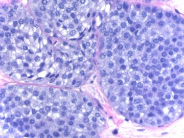
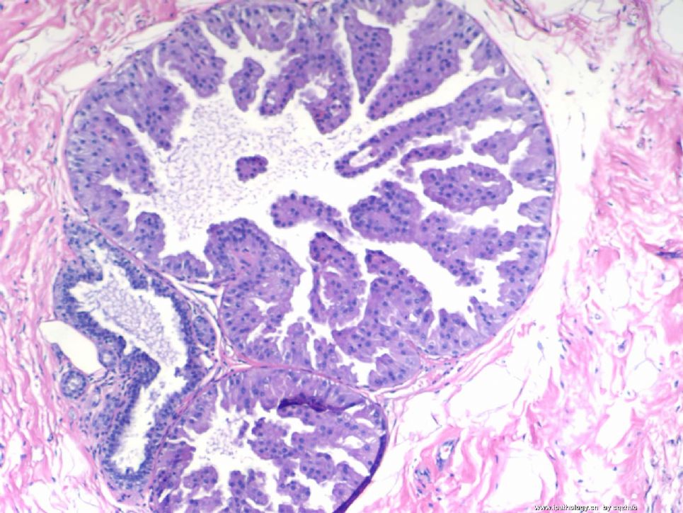
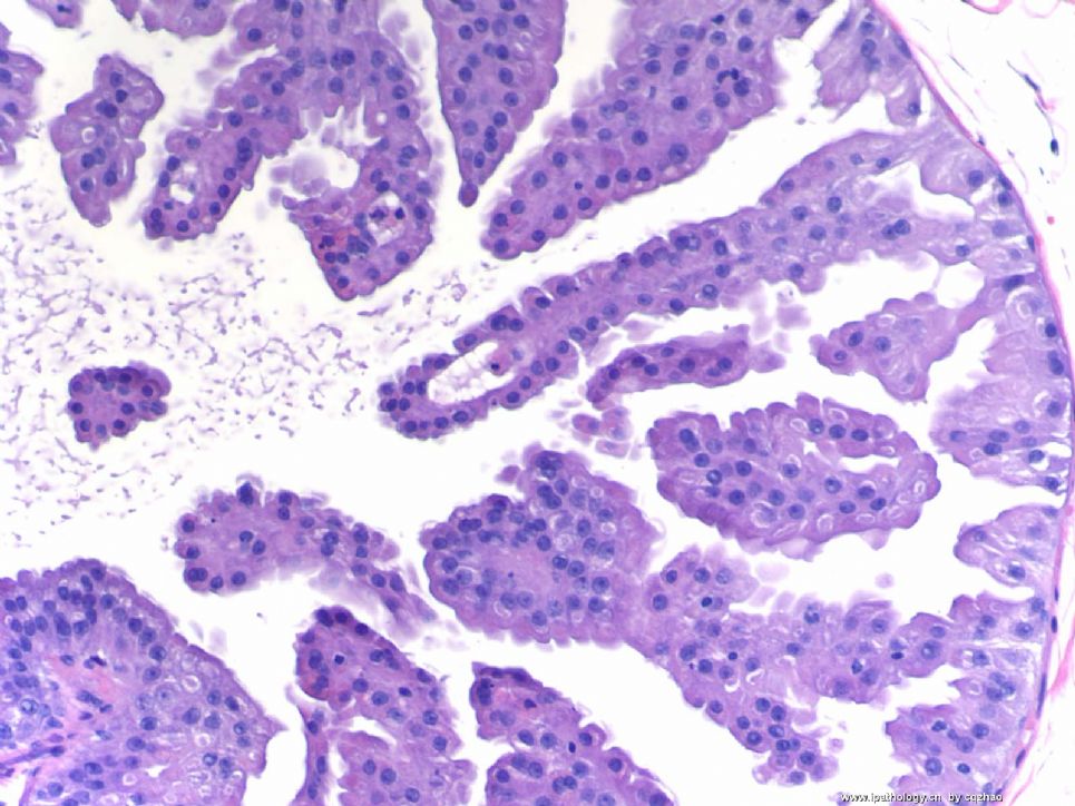
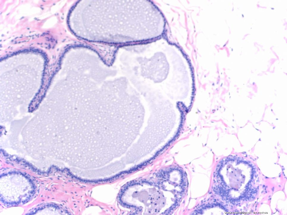
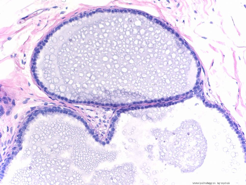
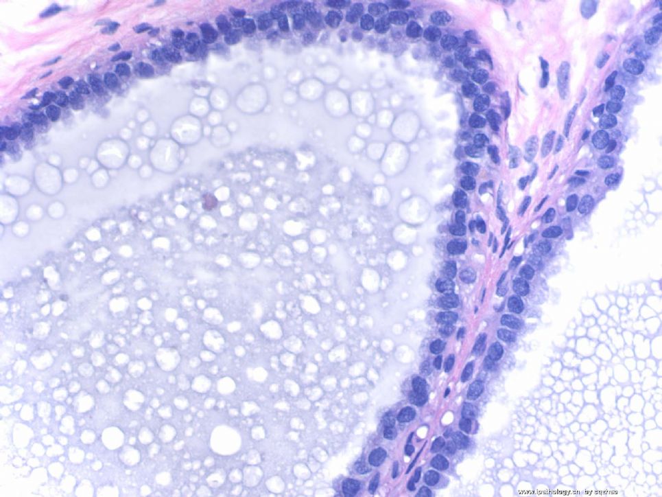
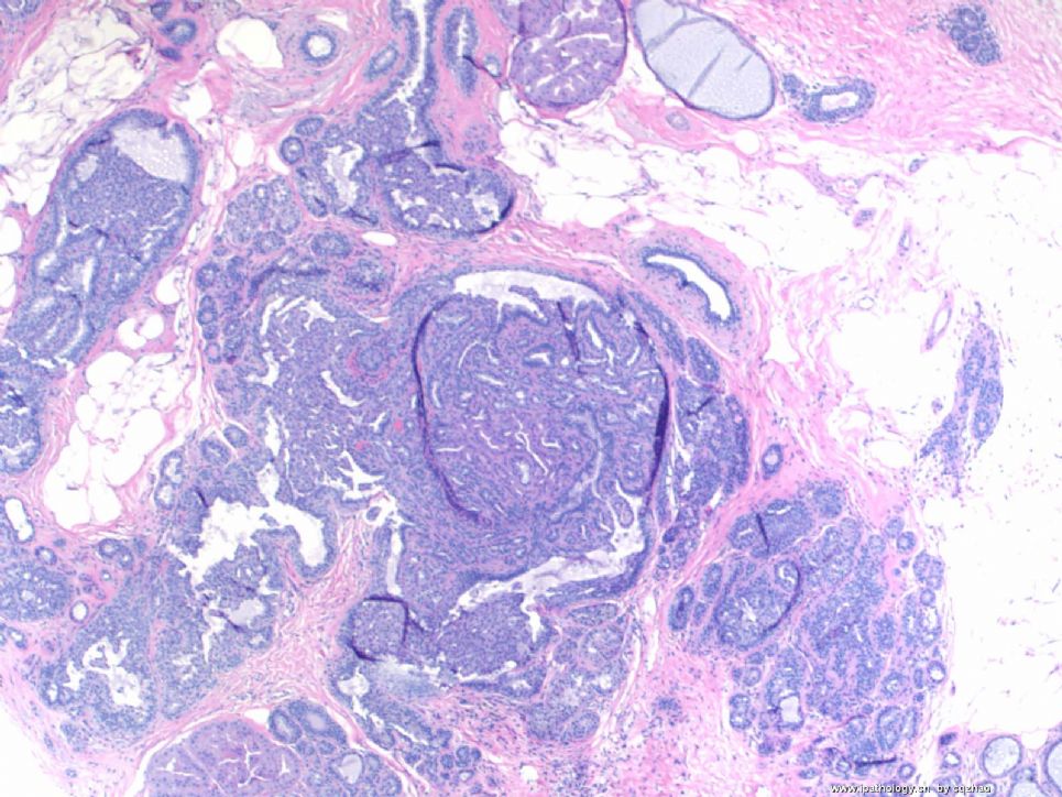
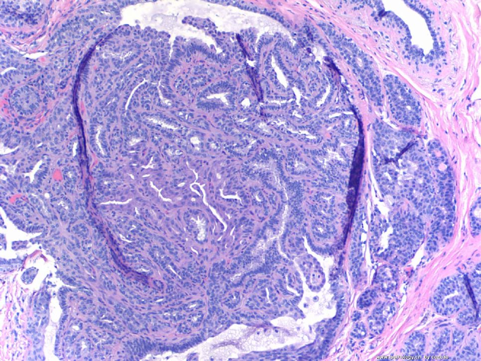
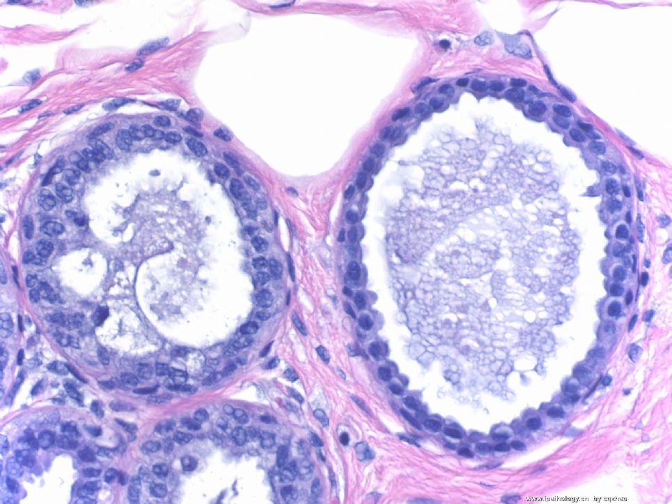
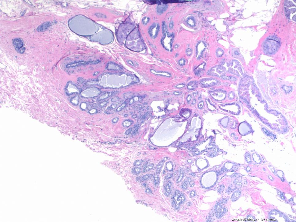
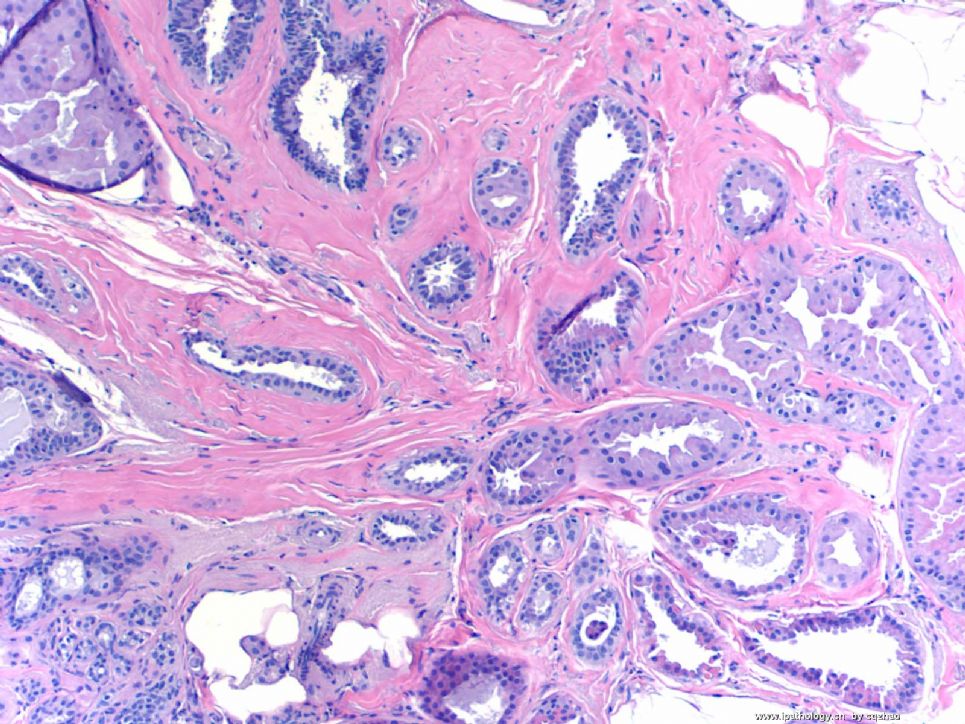
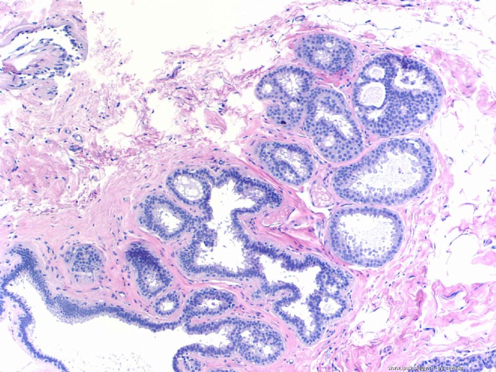
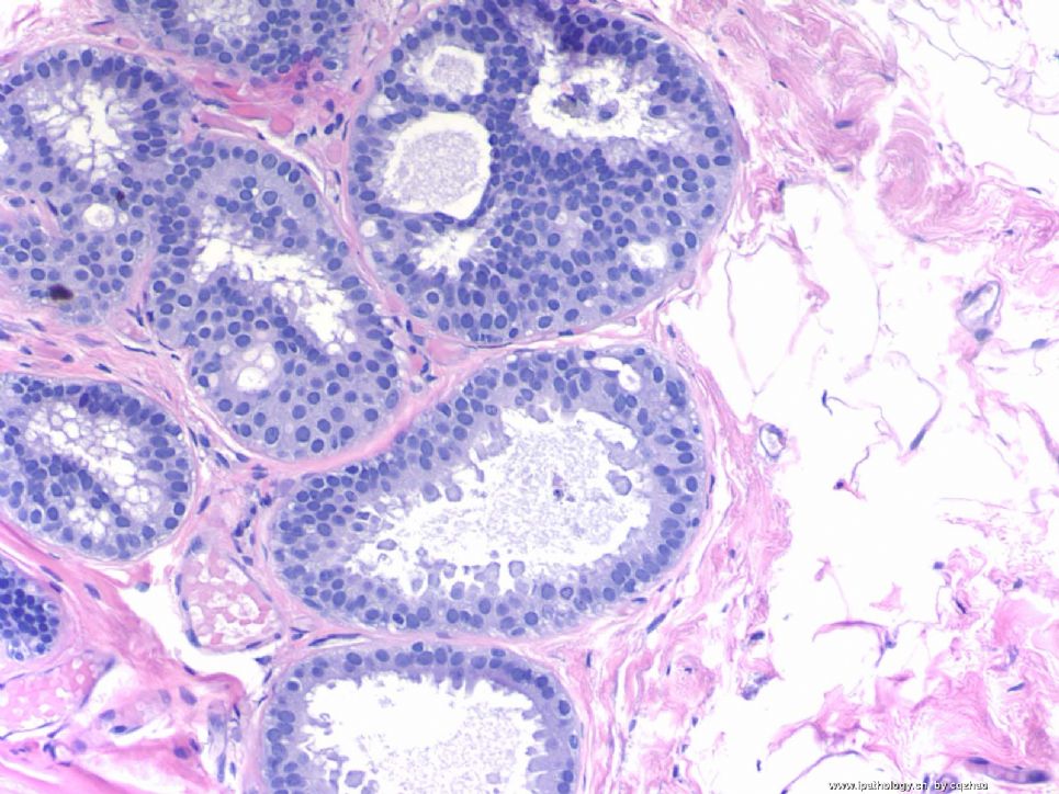
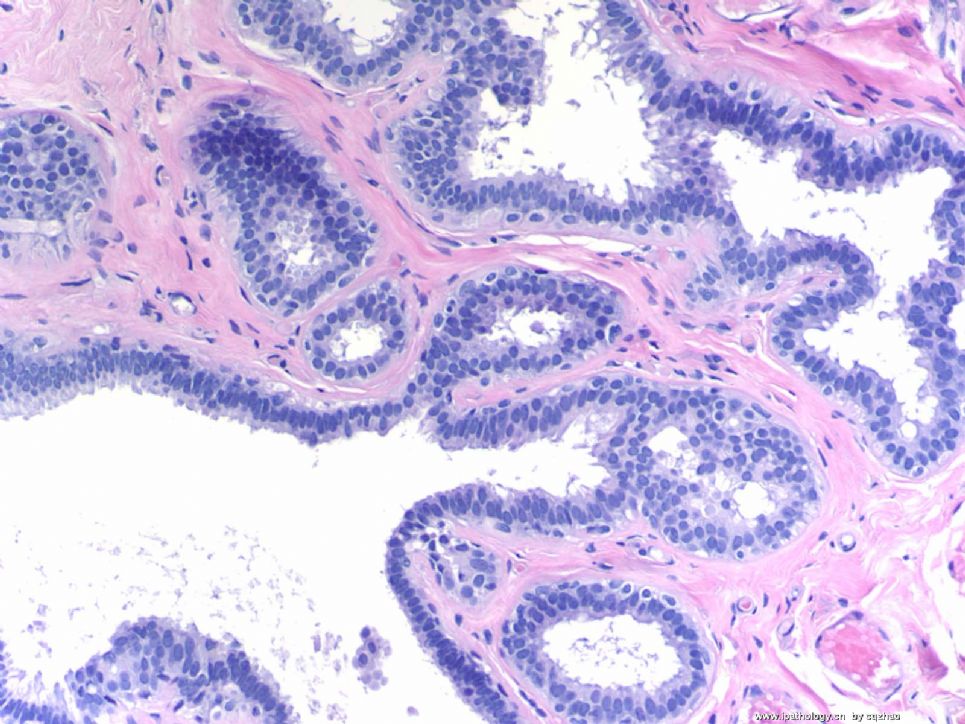
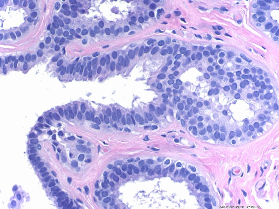
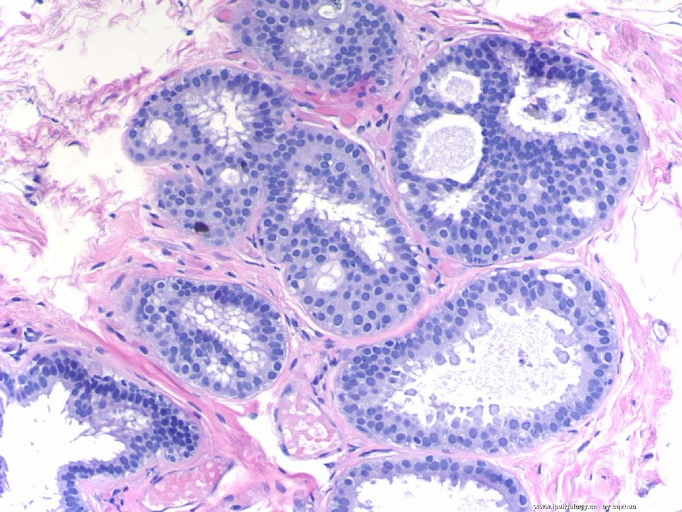
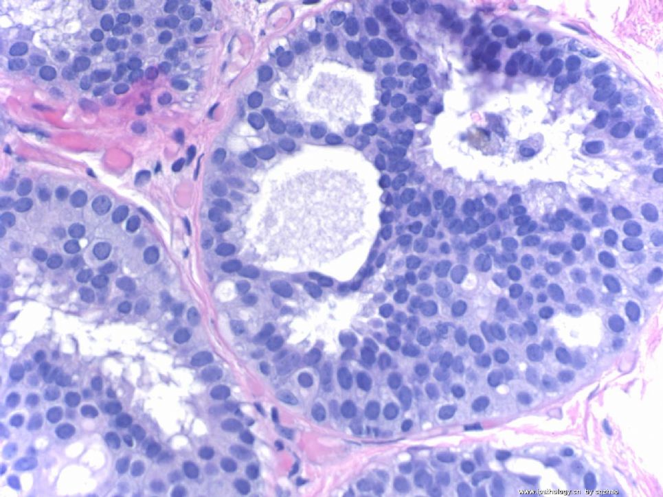
Breast core biopsy with 8 cores. There are multiple breast lesions. Try you diagnosis one by one.
Lesion 1 20x and 400x
-
本帖最后由 于 2009-03-19 23:29:00 编辑
相关帖子
- • 乳腺癌?
- • 女性 冰冻为乳腺浸润性导管癌,现切除标本,肿块旁组织
- • 女性 33岁 乳腺肿块
- • 乳腺包块
- • 乳腺两个相邻导管内的病变
- • 乳腺肿物
- • 38岁乳腺(新加HE切片)
- • 乳腺包块。33岁
- • 左乳肿块,协助诊断
- • 乳腺肿物求助
Breast pathology is complicated. We as pathologists need to recognize the cancers and all other borderline or benign lesions. There are several lesions in this breast core specimen. Please make your dx based on above orders, lesion 1, 2, 3,....
Thanks,
cz
| 以下是引用luolili在2009-2-18 14:22:00的发言:
此病变有以下特征: 1 导管上皮增生,伴有轻-中度非典型增生 2 导管上皮乳头状增生,伴大汗腺化生 3 导管囊性扩张伴分泌物潴留 4 导管内乳头状瘤 5 ?是导管良性病变 6 纤维囊性乳腺病 7 腺病,导管上皮增生,伴中度非典型增生。 新手上路,望各位老师多多指教。 |
1. Lobular carcinoma in situ.
2.atypical duct hyperplasia, papillary variant.
3.dilated duct.
4. papilloma.
5.columnar cell change
6.sclerosing adenosis
7.columnar cell lesions including columnar cell change, columnar cell hyperplasia,flat epithelial atypia(atypical duct hyperplasia?)
-
1. lobular neoplasia
2. nuclear grade 1 DCIS, apocrine type
3. flat epithelial atypia
4. florid ductal hyperplasia/intraductal papilloma
5. flat epithelial atypia and nuclear grade 1 DCIS
6. radial scar
7. a. columnar cell change, b.atypical coloumnar cell change, c. cribriform DCIS
Quickly review all of your interpretation. Most of them are reasonable. In this topic I will concentrate for the interpretaion of FEA, columnar cell change. If you are interested you can review the text book or related articles for some photos first. Then we can have some discussion together. Dr. Stuart, J. Schnitt from Beth Israel Deaconess Medical Center, Harward Medical School, Boston, MA did a lot of study in this area. In fact he raised some terms. Frequently speaking, I am often confused by the diagnosis of FEA in my clinical practice.
Hope more people share the diagnosis about my first 7 lesions from the same patient.
Thanks,
cz
