共4页/65条首页上一页1234下一页尾页
回复:65 阅读:9814
B1581Breast lesions cqz (12) (2-16-2009)

cqzhao 离线

帖子:5518
粉蓝豆:1071
经验:5755
注册时间:2008-09-29
加关注  |  发消息
楼主 发表于 2009-02-17 09:05|举报|关注(1)
浏览排序[ 顺序 逆序 楼主 支持 精彩 ]  快捷回复
姓    名: ××× 性别:   年龄:  
标本名称:  
简要病史:  
肉眼检查:  

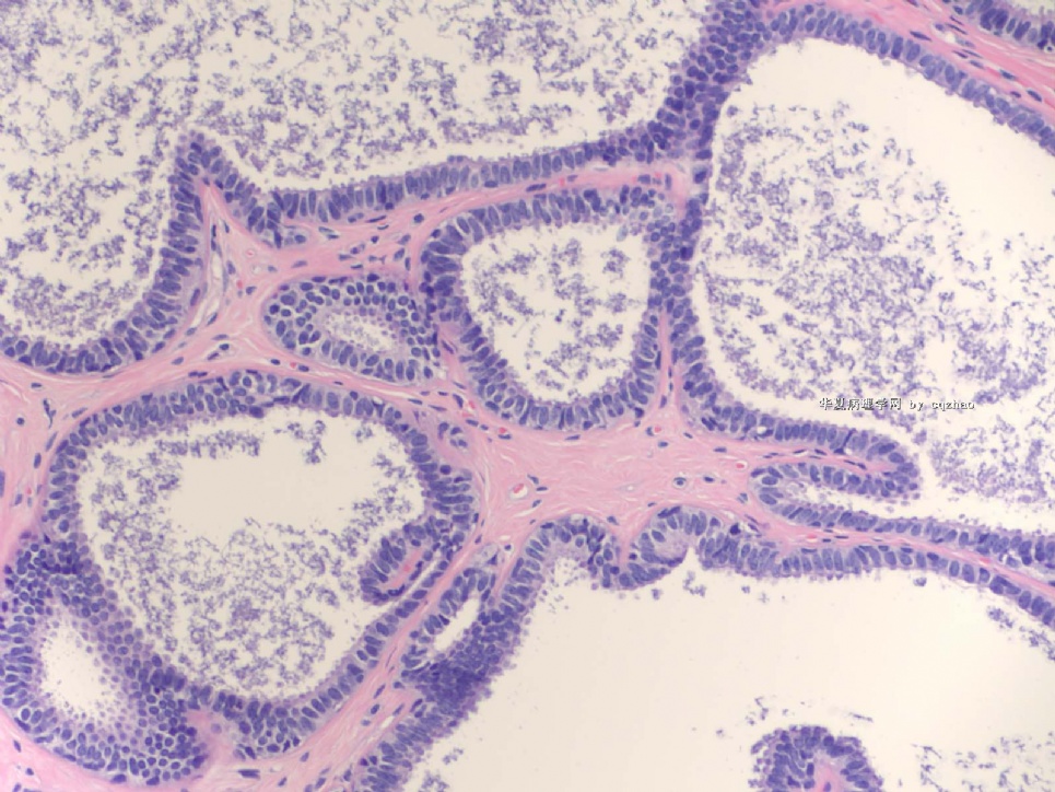
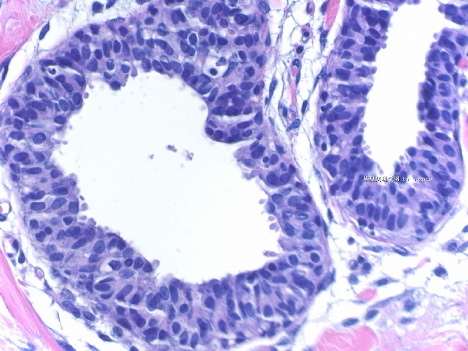
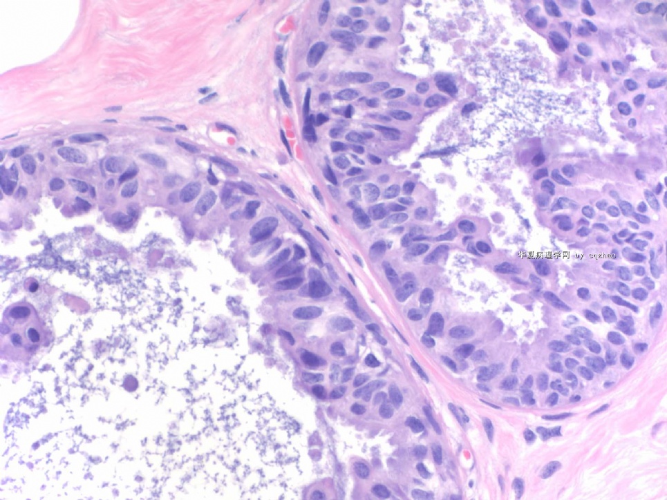
Breast core biopsy with 8 cores. There are multiple breast lesions. Try you diagnosis one by one.

Lesion 1 20x and 400x

  • Breast lesions cqz (12) (2-16-2009)图1
    图1
  • Breast lesions cqz (12) (2-16-2009)图2
    图2
标签:LCIS CCC FEA ADH 导管内乳头状肿瘤 放射状瘢痕
本帖最后由 于 2009-03-19 23:29:00 编辑
0
添加参考诊断
×参考诊断
  

Elizabeth 离线

帖子:59
粉蓝豆:1
经验:59
注册时间:2008-12-30
加关注  |  发消息
41 楼    发表于2009-03-14 09:23:00举报|引用
返回顶部 | 快捷回复
 Fig. 11.11. Intraductal carcinoma, flat (“clinging”) micropapillary type. A: The papillary structures in this cystically dilated duct contain fractured calcifications of the ossifying type typically associated with columnar cell lesions. B: Carcinoma cells with pleomorphic nuclei and a disorderly distribution line the duct and overlie the calcification. C: The flat carcinomatous epithelium displays apical apocrine-type cytoplasmic “snouts.” D,E: Another example of flat, “clinging” intraductal carcinoma

_ from Rosen Breast Pathology.   Compare with lesion 3, just for learning.

0
回复

cqzhao 离线

帖子:5518
粉蓝豆:1071
经验:5755
注册时间:2008-09-29
加关注  |  发消息
42 楼    发表于2009-03-14 10:36:00举报|引用
返回顶部 | 快捷回复
本帖最后由 于 2009-03-14 10:40:00 编辑

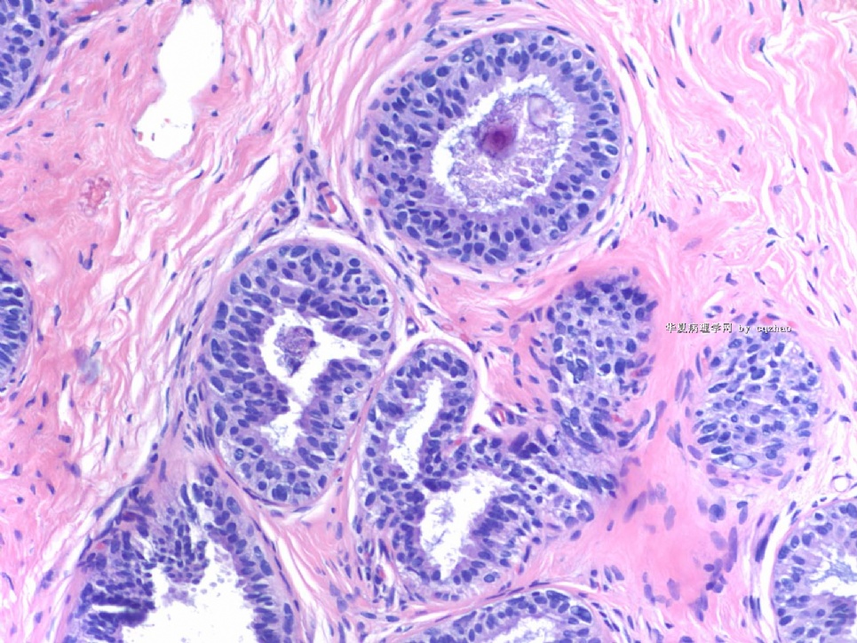
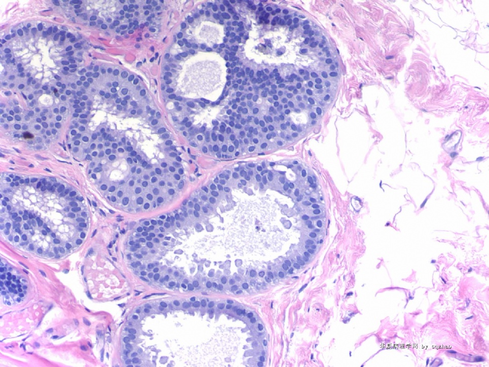
 Compared Rosen fig 11.11 (page 298 in the third edition) with the above lesion 3, we can appreciate the difference in term of cytology, uniform without atypia in lesion 3 and moderate cytologic atypia in Rosen's fig 11.11.

It is very good for the disagreement. I hope more pathologists join in the diacussion.

0
回复

cqzhao 离线

帖子:5518
粉蓝豆:1071
经验:5755
注册时间:2008-09-29
加关注  |  发消息
43 楼    发表于2009-03-14 10:52:00举报|引用
返回顶部 | 快捷回复
本帖最后由 于 2009-03-19 10:28:00 编辑  Peter Rosen is an excellent breast pathologist. His Book " Rosen's Breast Pathology in the best comprehensive book in the area of breast pathology. His calssification about columnar cell lesion is different from most others. He used the term columinar cell hyperplasia, mild, moderate, and severe atypia. I really do not like his concept about CCC. It is true that intraductal carcinoma, flat pattern, is present.  However, I fell reluctant to call the 5 photos Elizabeth took from Rosen's book as carcinoma based on the photos only.
0
回复

cqzhao 离线

帖子:5518
粉蓝豆:1071
经验:5755
注册时间:2008-09-29
加关注  |  发消息
44 楼    发表于2009-03-18 05:09:00举报|引用
返回顶部 | 快捷回复

 Thank Dr. Chiang' s interpretation.

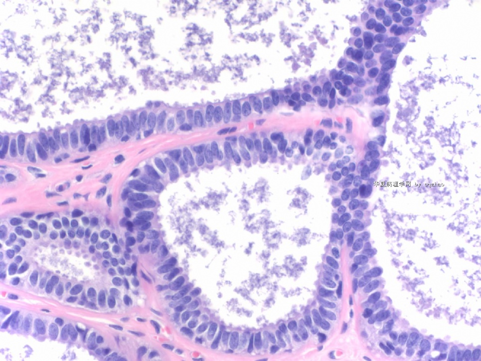
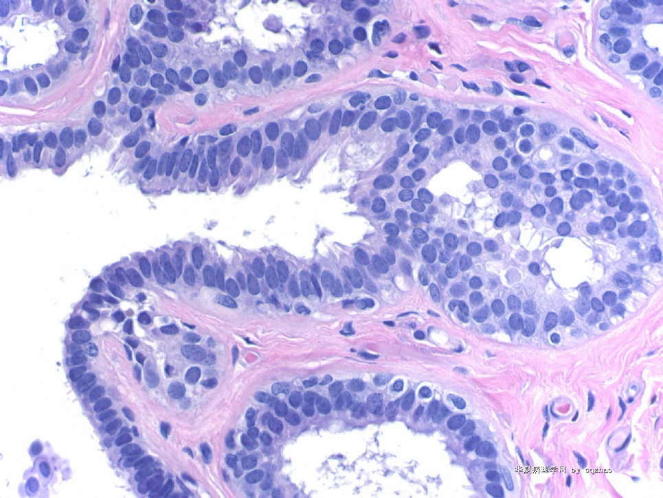
Lesion 2: Apocrine metaplasia or change with micropapaillary or papillary pattern. The nuclei are very uniform, small, evenly spaced, located the botton of the cells. I do not appreciate any cytologic atypia in this case. Cystic papillary apocrine changes are commonly seen in the breast pathology specimens. Generally people will ignorred the lesion even though mild cytologic atypia is present in the apocrine change. True atypical apocrine metaplasia is rare.

0
回复

有福不在忙 离线

帖子:2161
粉蓝豆:42
经验:3448
注册时间:2008-11-22
加关注  |  发消息
45 楼    发表于2009-03-18 22:36:00举报|引用
返回顶部 | 快捷回复
 学习了。
0
回复
signature
有福不在忙

cqzhao 离线

帖子:5518
粉蓝豆:1071
经验:5755
注册时间:2008-09-29
加关注  |  发消息
46 楼    发表于2009-03-19 10:37:00举报|引用
返回顶部 | 快捷回复
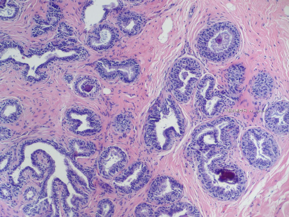
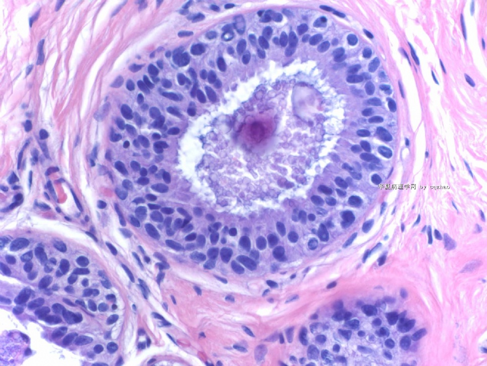
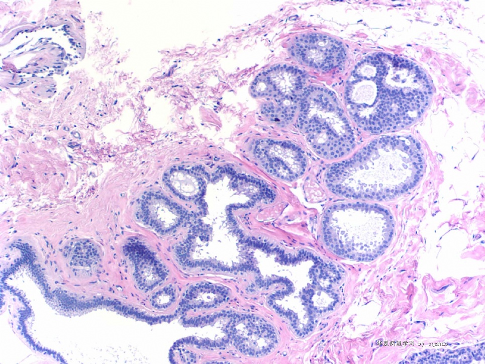
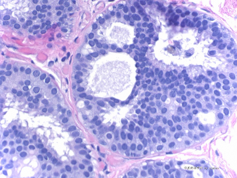
 Lesion 3: 导管囊性扩张伴分泌物潴留 is a good call. If you call CCC it is ok. Anyway I cannot appreciate atypia here. It is not a FEA or carcinoma in situ. Compared with the photos on 41 floor we will know what is carcinoma, flat type, even though these figures are not very typical ones.
0
回复

abin 离线

帖子:7383
粉蓝豆:6013
经验:11754
注册时间:2006-10-08
加关注  |  发消息
47 楼    发表于2009-03-19 23:36:00举报|引用
返回顶部 | 快捷回复

 可惜我迟到了。也写下自己的意见:

1 DCIS
2 CCC
3 纤维囊性乳腺病伴CCC及分泌
4 周围型乳头状瘤
5 CCC or DCIS
6 硬化性腺病+CCC
7 DCIS+CCC+纤维囊性乳腺病

0
回复
signature

华夏病理/粉蓝医疗

为基层医院病理科提供全面解决方案,

努力让人人享有便捷准确可靠的病理诊断服务。


cqzhao 离线

帖子:5518
粉蓝豆:1071
经验:5755
注册时间:2008-09-29
加关注  |  发消息
48 楼    发表于2009-03-20 02:19:00举报|引用
返回顶部 | 快捷回复
 lesion 1. IHC confirmed it is a lobular lesion. LCIS.
0
回复

cqzhao 离线

帖子:5518
粉蓝豆:1071
经验:5755
注册时间:2008-09-29
加关注  |  发消息
49 楼    发表于2009-03-20 02:23:00举报|引用
返回顶部 | 快捷回复
本帖最后由 于 2009-03-20 02:28:00 编辑
以下是引用cqzhao在2009-2-17 9:21:00的发言:

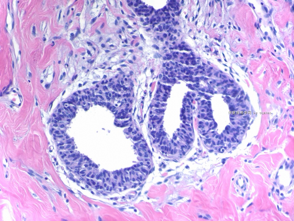
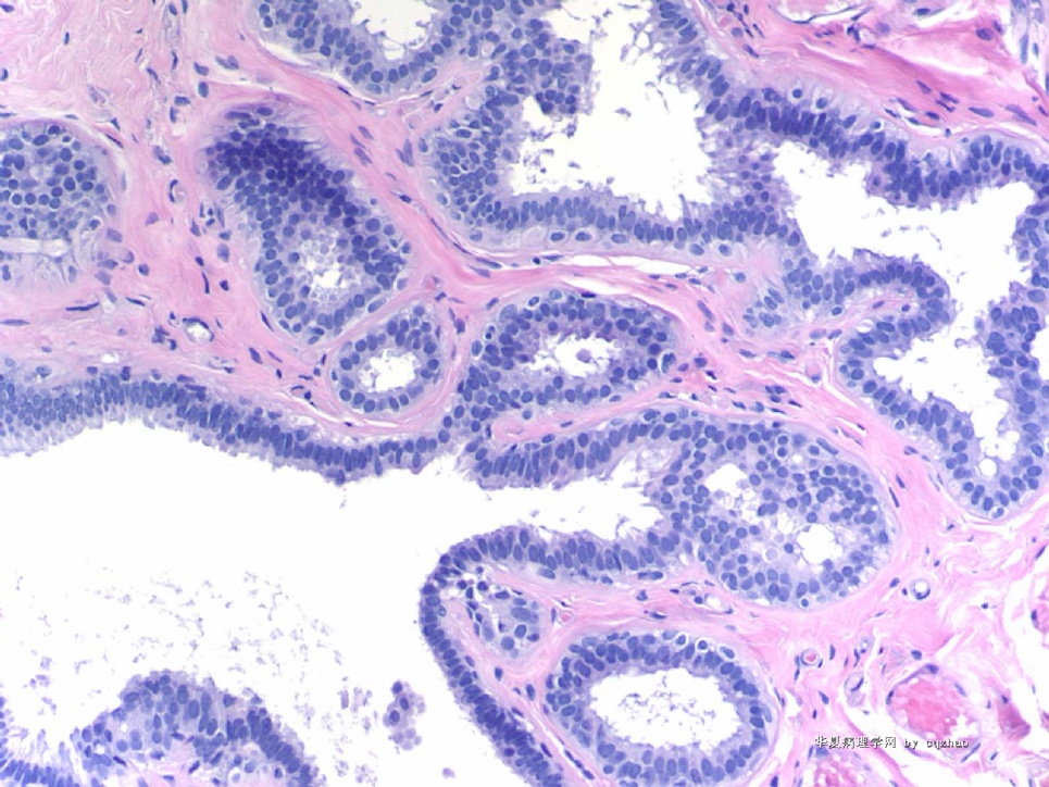
 Lesion 7

Fig 1 100x

Fig 2-5 200x

Fig 6 400x

Hope you can appreciate three diagnoses (or three terms) in this focal area.

There are some different oppinion about this lesion. Pasted the figs again.

CCC, FEA, ADH.

Question is if the dianosis of DCIS should be rendered.

Is the severe lesion in this area ADH or DCIS?

Please share your oppinion and the reasons.

Thanks, cz


名称:图1
描述:图1

名称:图2
描述:图2

名称:图3
描述:图3

名称:图4
描述:图4

名称:图5
描述:图5

名称:图6
描述:图6
0
回复

student 离线

帖子:16
粉蓝豆:1
经验:27
注册时间:2008-06-30
加关注  |  发消息
50 楼    发表于2009-03-21 20:24:00举报|引用
返回顶部 | 快捷回复

 再次学习了,赞同Dr.Zhao

Lesion 7不足DCIS,称为ADH比较合适。

Lesion 1更像LCIS。而且有IHC证实。

0
回复

天山望月 离线

帖子:4902
粉蓝豆:83
经验:5092
注册时间:2007-02-03
加关注  |  发消息
51 楼    发表于2009-03-22 10:52:00举报|引用
返回顶部 | 快捷回复
本帖最后由 于 2009-03-23 12:41:00 编辑

 谢谢Dr.zhao!如此丰盛的大餐!

先来回答问题,不知得多少分,后再阅读大家讨论学习。

1、LCIS,需IHC标记P120,E-Ca排除导管癌累及小叶。

2、乳头状大汗腺化生。

3、柱状上皮病变?分泌性肿瘤或病变???????????

4、导管内乳头状瘤。

5、柱状上皮病变。

6、纤维腺病伴大汗腺化生。Check the photo again

7、腺病,UDH。Are you sure?

0
回复
signature
广州金域病理

cqzhao 离线

帖子:5518
粉蓝豆:1071
经验:5755
注册时间:2008-09-29
加关注  |  发消息
52 楼    发表于2009-03-23 12:23:00举报|引用
返回顶部 | 快捷回复
 Lesion 7 demostrates focal ductal epithelial cell proliferation with uniform size in the back ground of CCC or focal FEA. It is reasonable for diagnosis of ADH. The lesion does not meet  the DCIS  criteria  by 2-3 mm or 2-3 ducts. Hope your guys will agree.
0
回复

cqzhao 离线

帖子:5518
粉蓝豆:1071
经验:5755
注册时间:2008-09-29
加关注  |  发消息
53 楼    发表于2009-03-23 12:27:00举报|引用
返回顶部 | 快捷回复
本帖最后由 于 2009-03-23 12:30:00 编辑

 I will show you more photos.

Case 1

100x

200x

400x


名称:图1
描述:图1

名称:图2
描述:图2

名称:图3
描述:图3
0
回复

cqzhao 离线

帖子:5518
粉蓝豆:1071
经验:5755
注册时间:2008-09-29
加关注  |  发消息
54 楼    发表于2009-03-23 12:31:00举报|引用
返回顶部 | 快捷回复
本帖最后由 于 2009-03-23 12:32:00 编辑

 Case 2

200x

400x


名称:图1
描述:图1

名称:图2
描述:图2
0
回复

cqzhao 离线

帖子:5518
粉蓝豆:1071
经验:5755
注册时间:2008-09-29
加关注  |  发消息
55 楼    发表于2009-03-23 12:32:00举报|引用
返回顶部 | 快捷回复
本帖最后由 于 2009-03-23 12:34:00 编辑

 Case 3

100x

200x

300x


名称:图1
描述:图1

名称:图2
描述:图2

名称:图3
描述:图3
0
回复

cqzhao 离线

帖子:5518
粉蓝豆:1071
经验:5755
注册时间:2008-09-29
加关注  |  发消息
56 楼    发表于2009-03-23 12:34:00举报|引用
返回顶部 | 快捷回复
本帖最后由 于 2009-03-23 12:36:00 编辑

 case 4 400x

case 5 400x

case 6 400x


名称:图1
描述:图1

名称:图2
描述:图2

名称:图3
描述:图3
0
回复

cqzhao 离线

帖子:5518
粉蓝豆:1071
经验:5755
注册时间:2008-09-29
加关注  |  发消息
57 楼    发表于2009-03-23 12:38:00举报|引用
返回顶部 | 快捷回复
 Plese write your interpretation from case 1 to case 6. Thanks, cz
0
回复

天山望月 离线

帖子:4902
粉蓝豆:83
经验:5092
注册时间:2007-02-03
加关注  |  发消息
58 楼    发表于2009-03-23 22:05:00举报|引用
返回顶部 | 快捷回复
本帖最后由 于 2009-03-23 22:21:00 编辑

 先回52楼Dr.zhao的问题:

重新阅图,图7,我不敢确定,看了大家积极的讨论和赵博士的答案,明白了。

那天急着往回赶,没来得及看后面的讨论。

0
回复
signature
广州金域病理

天山望月 离线

帖子:4902
粉蓝豆:83
经验:5092
注册时间:2007-02-03
加关注  |  发消息
59 楼    发表于2009-03-23 22:19:00举报|引用
返回顶部 | 快捷回复
本帖最后由 于 2009-03-23 22:20:00 编辑

 回复54楼后病例:

1、柱状上皮病变,细胞有些拥挤,但异型性不明显。

2、3、非典型性的柱状上皮病变,(可以称为平坦型DCIS吗?)

4、DCIS.

5、平坦型的DCIS。

6、非典型的柱状上皮病变。

呵呵,没信心了,好像都是一个主题,赶快回去查资料,并敬等打分。

0
回复
signature
广州金域病理

cqzhao 离线

帖子:5518
粉蓝豆:1071
经验:5755
注册时间:2008-09-29
加关注  |  发消息
60 楼    发表于2009-04-05 03:48:00举报|引用
返回顶部 | 快捷回复
 Just check to notice only 天山望月 writing answers for the last four cases. They are too easy for your guys or you do not like these kinds of cases. In fact they are very hot area in current breast pathology. Hope more people know these lesions.
0
回复
回复:65 阅读:9814
共4页/65条首页上一页1234下一页尾页
【免责声明】讨论内容仅作学术交流之用,不作为诊疗依据,由此而引起的法律问题作者及本站不承担任何责任。
快速回复
进入高级回复
您最多可输入10000个汉字,按 "Ctrl" + "Enter" 直接发送
搜索回复/乘电梯 ×
按内容
按会员
乘电梯
合作伙伴
友情链接