| 图片: | |
|---|---|
| 名称: | |
| 描述: | |
- Breast lobular lesions and stains ( cqz 7)
-
本帖最后由 于 2008-12-14 14:52:00 编辑
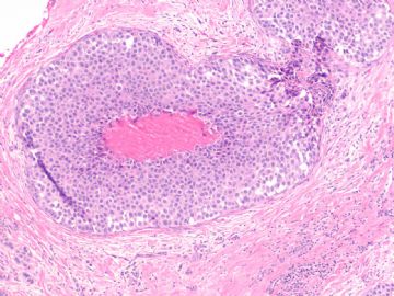
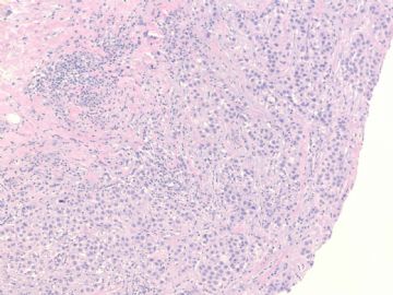
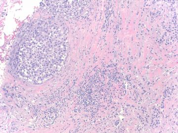
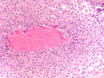
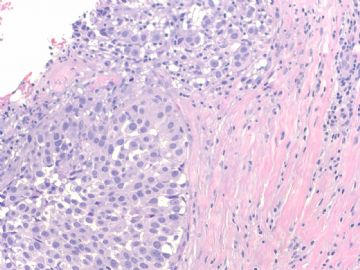
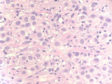
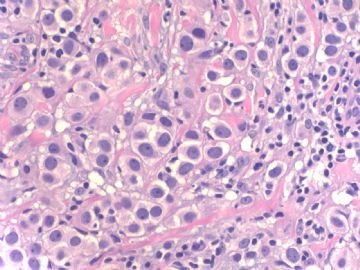
pleomorphic variant?
Some invasive lobular carcinomas consist entirely or in part of cells larger than cells in classical invasive lobular carcinoma with relatively abundant, eosinophilic cytoplasm (Fig. 32.17). The nucleus in some examples is hyperchromatic and eccentric with a distinct nucleolus creating a plasmacytoid appearance (Fig. 32.18). These cells have been referred to variously as myoid (12), histiocytoid (77-79), and pleomorphic lobular carcinoma (80,81) (Figs. 32.18-19). __from Rosen's Breat Pathology (P700)

华夏病理/粉蓝医疗
为基层医院病理科提供全面解决方案,
努力让人人享有便捷准确可靠的病理诊断服务。
| 以下是引用cqzhao在2008-12-14 0:25:00的发言:
People aged 40 above are proffesors or experts already. They do not need to learn. |
 Maybe they learn by other ways.

华夏病理/粉蓝医疗
为基层医院病理科提供全面解决方案,
努力让人人享有便捷准确可靠的病理诊断服务。
| 以下是引用天山望月在2008-12-13 23:21:00的发言:
|
People aged 40 above are proffesors or experts already. They do not need to learn.
-
本帖最后由 于 2008-12-14 14:06:00 编辑
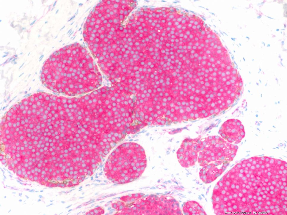
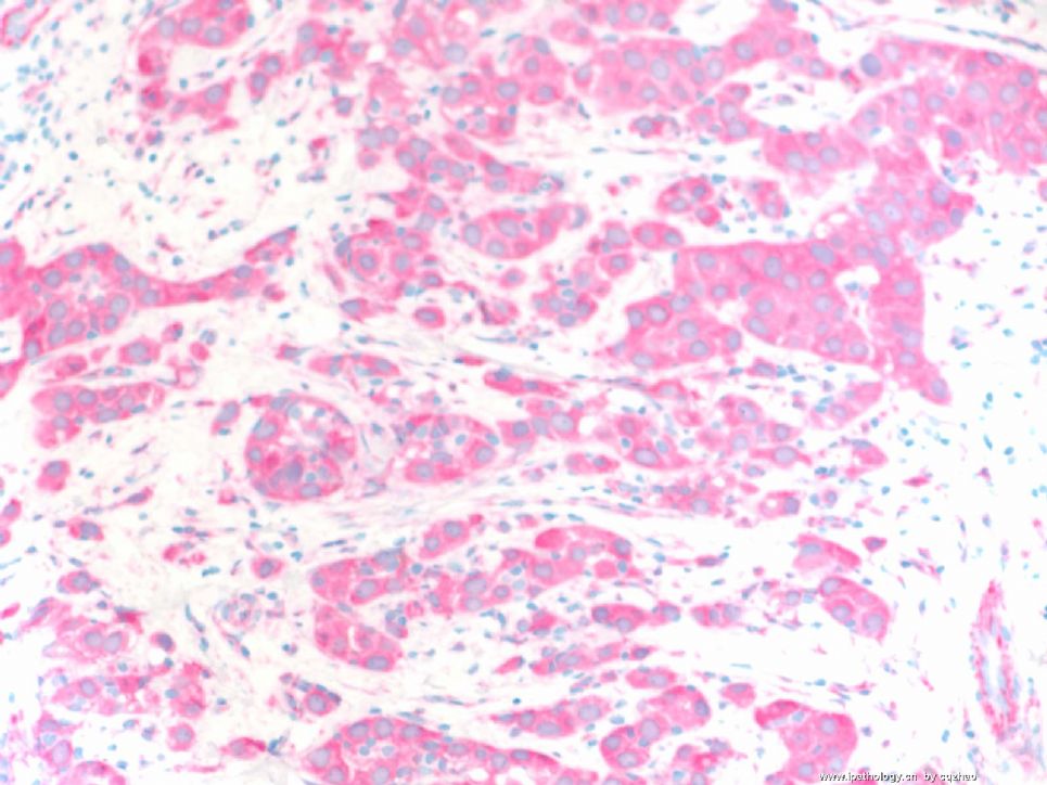
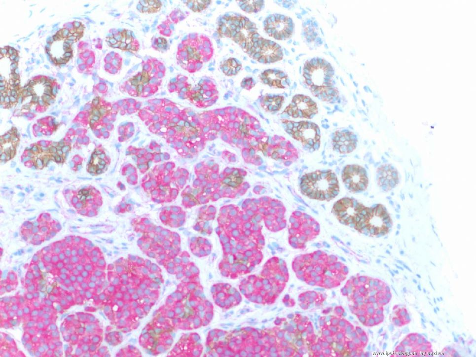
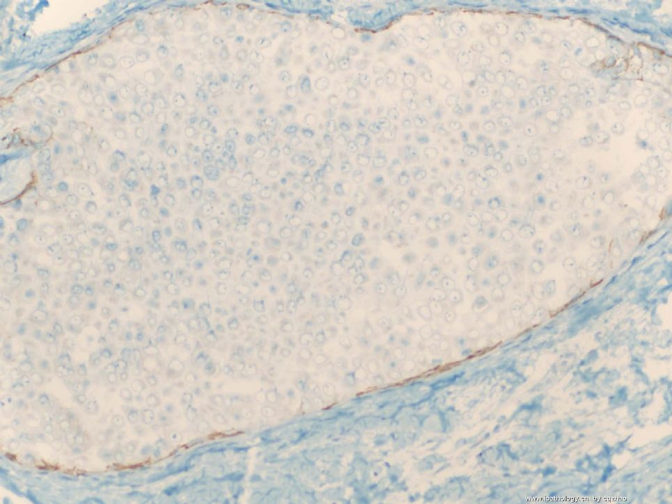
Dual stains: anti-ecad labeling with brown color, anti-p120 with pink color, mixed stain, one procedure.
F 1 for insi tu ca
F2 for invasive ca
F3 in situ and normal ducts.
E-cad membrane stain (brown)
P-120 cytoplasmic stain (pink)
Lobular lesion will not see brown color and only pick.
Fig 3: some normal ducts show brown membrane stain and no pink color.
So you know the nature of the lesions.
Lobular carcinoma in situ and invasive lobular carcinoma.
Now, what type of invasive lobular carcinoma is it?
abin译:
免疫组化双标记:抗E-cadherin棕色,抗p120粉红色。
图1 原位癌
图2 浸润癌
图3 原位癌和正常导管。E-cadherin呈膜阳性(棕色),P-120呈胞浆阳性(粉红色)
小叶病变不见棕色,只有粉红色。正常导管示棕色的膜染色,没有粉红色。
因此你明白病变的性质了:小叶原位癌和浸润性小叶癌。
那么,是什么亚型的浸润性小叶癌呢?
名称:图1
描述:图1
名称:图2
描述:图2
名称:图3
描述:图3
-
本帖最后由 于 2008-12-14 14:51:00 编辑
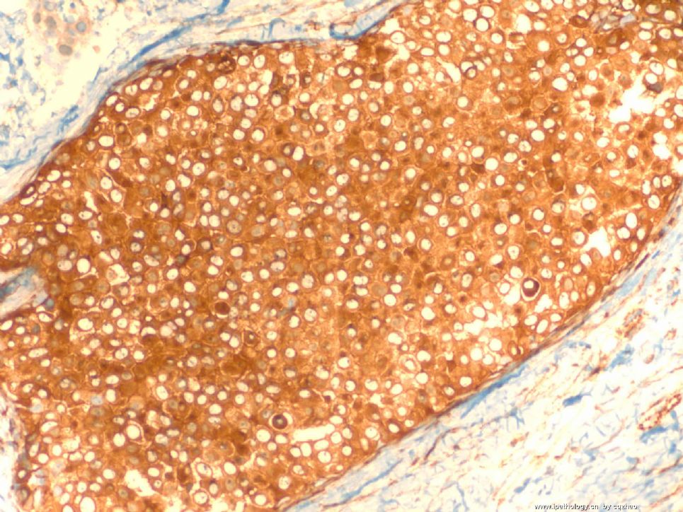
Photos 200x
F1 E-cadherine for insitu ca 图1 原位癌E-cadherin染色
F2 E-cadherine for invasive 图2 浸润癌E-cadherin染色
F3 P120 for insitu ca 图3 原位癌P120染色
F4. P120 for invasive ca 图4 浸润癌P120染色
Ductal ca (or normal ducts): memberane stain for both e-cad and p120
Lobular lesion: Absence of stain for E-cad and strong cytoplasmic stain for p120.
导管癌(或正常导管): E-cadherin和p120均呈膜阳性。
小叶病变:E-cadherin不着色,p120胞浆强阳性。
(abin译)
名称:图1
描述:图1

名称:图2
描述:图2
名称:图3
描述:图3

名称:图4
描述:图4
| 以下是引用cqzhao在2008-12-11 2:00:00的发言:
Good analysis. H&E slides are good enough to know the of insitu and invasive carcinoma. The key for this case is the nature of the tumor. I will take some photos when I have time. You can continue to guess. |
分析的好,HE切片足以识别原位癌和浸润癌,这个病例的关键是肿瘤的特性,有时间时,我会采一些图片,
大家继续讨论
闲来看云译
-
stevenshen 离线
- 帖子:343
- 粉蓝豆:2
- 经验:343
- 注册时间:2008-06-03
- 加关注 | 发消息
-
本帖最后由 于 2008-12-10 20:00:00 编辑
It is possible that it is invasive ductal carcinoma and solid DCIS; I would guess that it is LCIS with invasive lobular carcinoma (pleomorphic type). Thanks. Looking forward to hearing the discussion and final answer.
(abin译:这例可能是浸润性导管癌和实性DCIS。我猜也可能是LCIS伴浸润性小叶癌(多形性亚型)。谢谢。期待听到讨论和最终结果。)
-
wangxf_1997 离线
- 帖子:67
- 粉蓝豆:210
- 经验:77
- 注册时间:2008-08-22
- 加关注 | 发消息
