| 图片: | |
|---|---|
| 名称: | |
| 描述: | |
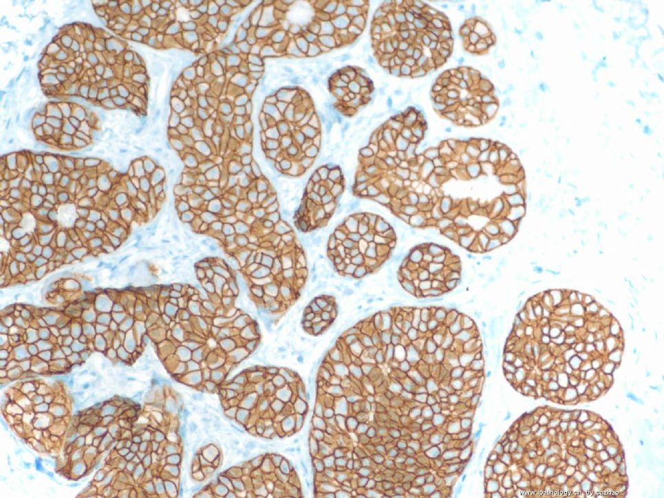
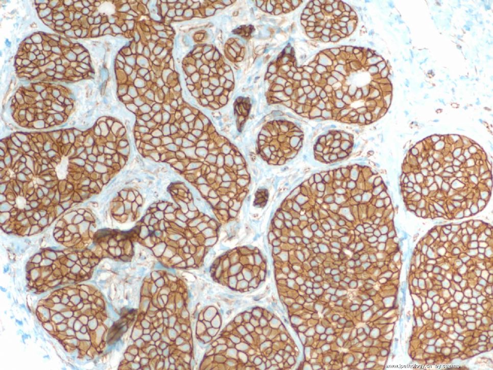
- Breast lobular lesions and stains ( cqz 7)
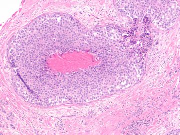
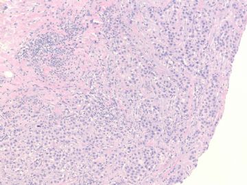
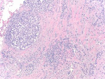
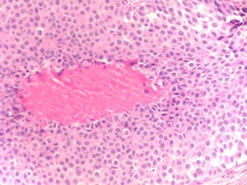
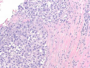
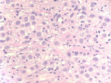
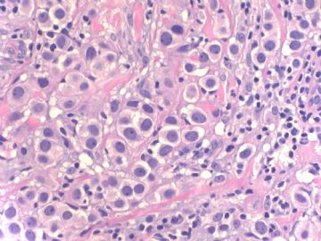
The same as most of you I first considered it was lobular carcinoma in situ in this core biopsy specimen. However, IHC results indicated it was a not lobular lesion. Now we know there are three possibilities, normal hyperplasia, ADH, and DCIS. Seeing the H&E slides more carefully, we know it is definitely abnormal. Generally the morphologic features of ADH are not like this. So only one possibility is DCIS with lobular extension or DCIS involving acini of lobules. If the specimens contain DCIS and DCIS with lobular extension, it is relatively easy to recongnize. If only lobular extension of DCIS is present in the specimen, it is difficult to make the diagnosis. But we should consider the possibility. Most of you did not consider the diagnosis. The reasons are you only see the photos, not true slides. Also the photos are not in the high power.
I reported lobular extension of intraductal carcinoma. The women had segmental mastectomy. The mastectomy specimen showed the DCIS closely to the previous core biopsy area.
This is for this case. Thanks.


 )。
)。
 the lesion is not lobular lesion?Looking forward for the final diagnosis.