| 图片: | |
|---|---|
| 名称: | |
| 描述: | |
- Breast lobular lesions and stains ( cqz 7)
-
本帖最后由 于 2008-12-14 14:06:00 编辑
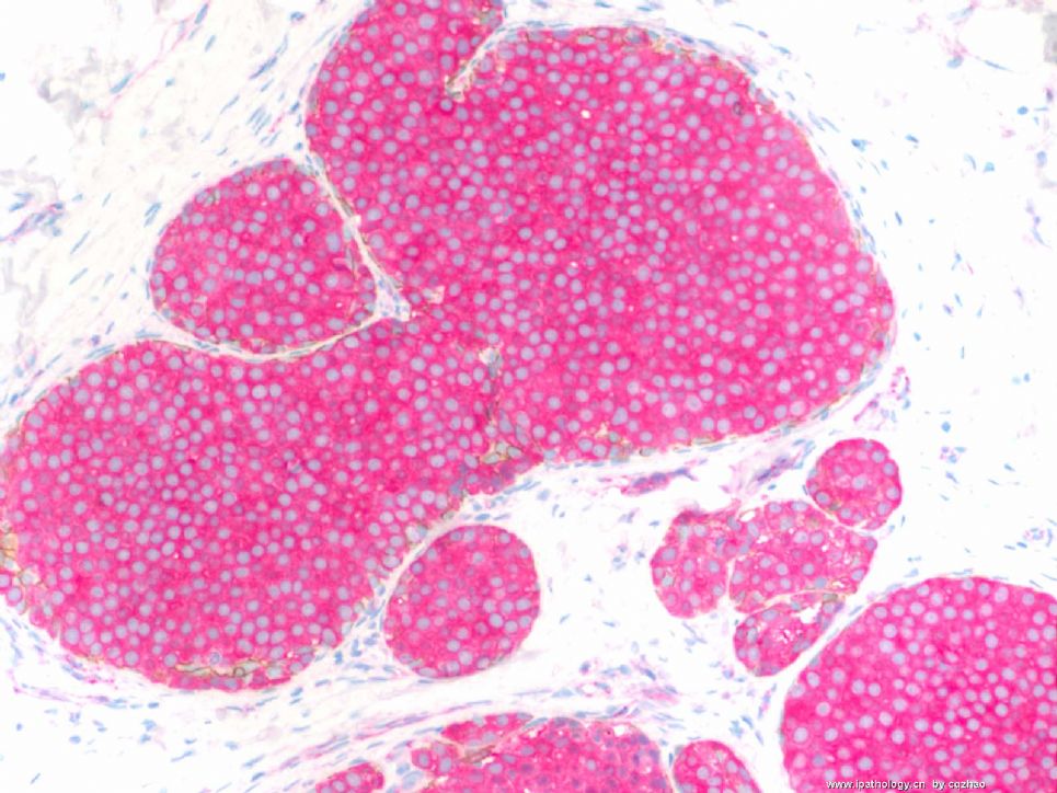
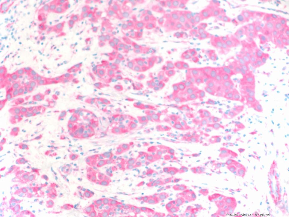
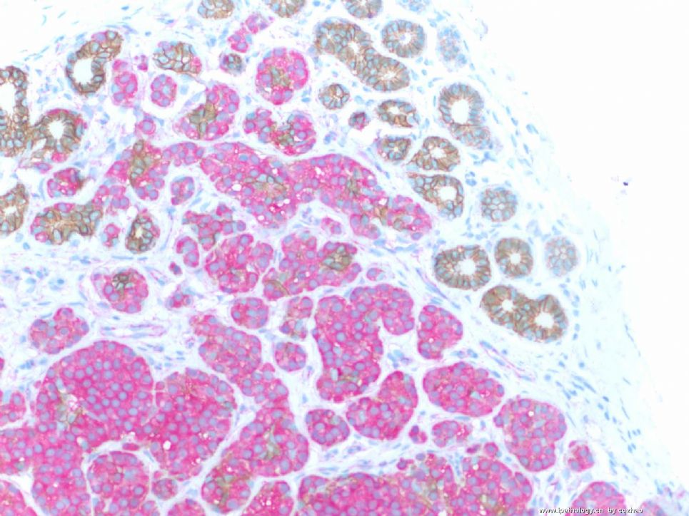
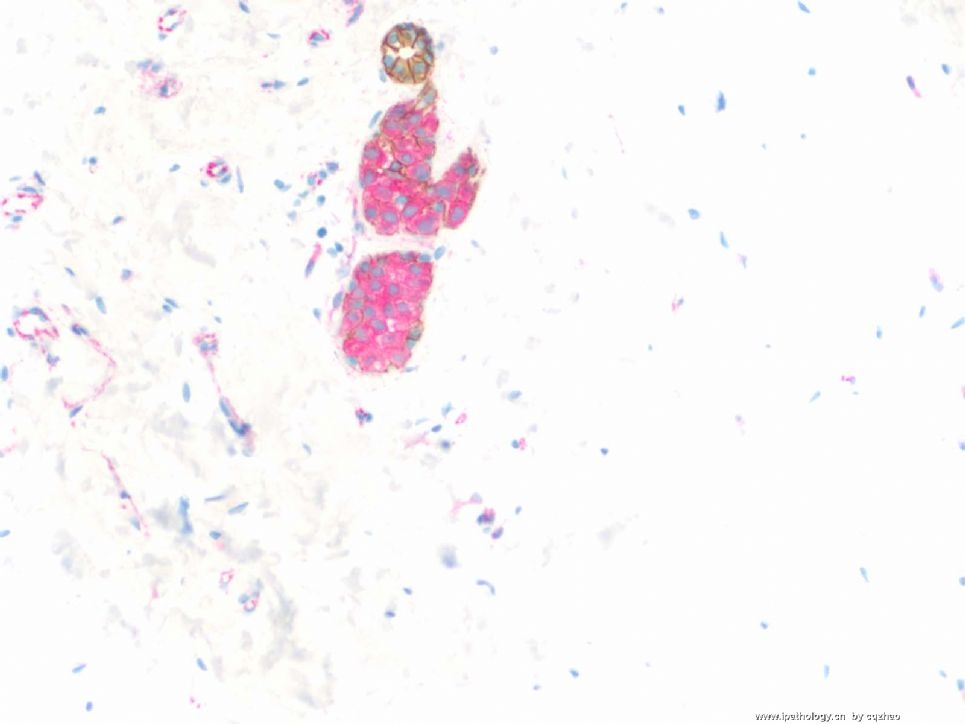
Dual stains: anti-ecad labeling with brown color, anti-p120 with pink color, mixed stain, one procedure.
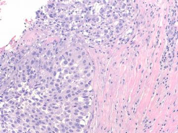
F 1 for insi tu ca
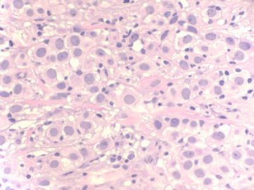
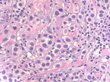
F2 for invasive ca
F3 in situ and normal ducts.
E-cad membrane stain (brown)
P-120 cytoplasmic stain (pink)
Lobular lesion will not see brown color and only pick.
Fig 3: some normal ducts show brown membrane stain and no pink color.
So you know the nature of the lesions.
Lobular carcinoma in situ and invasive lobular carcinoma.
Now, what type of invasive lobular carcinoma is it?
abin译:
免疫组化双标记:抗E-cadherin棕色,抗p120粉红色。
图1 原位癌
图2 浸润癌
图3 原位癌和正常导管。E-cadherin呈膜阳性(棕色),P-120呈胞浆阳性(粉红色)
小叶病变不见棕色,只有粉红色。正常导管示棕色的膜染色,没有粉红色。
因此你明白病变的性质了:小叶原位癌和浸润性小叶癌。
那么,是什么亚型的浸润性小叶癌呢?
名称:图1
描述:图1
名称:图2
描述:图2
名称:图3
描述:图3
| 以下是引用天山望月在2008-12-13 23:21:00的发言:
|
People aged 40 above are proffesors or experts already. They do not need to learn.
| 以下是引用cqzhao在2008-12-14 0:25:00的发言:
People aged 40 above are proffesors or experts already. They do not need to learn. |
 Maybe they learn by other ways.

华夏病理/粉蓝医疗
为基层医院病理科提供全面解决方案,
努力让人人享有便捷准确可靠的病理诊断服务。
-
本帖最后由 于 2008-12-14 14:52:00 编辑
pleomorphic variant?
Some invasive lobular carcinomas consist entirely or in part of cells larger than cells in classical invasive lobular carcinoma with relatively abundant, eosinophilic cytoplasm (Fig. 32.17). The nucleus in some examples is hyperchromatic and eccentric with a distinct nucleolus creating a plasmacytoid appearance (Fig. 32.18). These cells have been referred to variously as myoid (12), histiocytoid (77-79), and pleomorphic lobular carcinoma (80,81) (Figs. 32.18-19). __from Rosen's Breat Pathology (P700)

华夏病理/粉蓝医疗
为基层医院病理科提供全面解决方案,
努力让人人享有便捷准确可靠的病理诊断服务。
| 以下是引用cqzhao在2008-12-14 2:46:00的发言:
Now, what type of invasive lobular carcinoma is it? 那么,是什么亚型的浸润性小叶癌呢? |
先复习浸润性小叶癌的分型,再回答问题。
先描述一下浸润性小叶癌(ILC)的细胞形态特点:
A、经典型的细胞特点:多为黏附性差(即松散)的圆形卵圆形小细胞,浆少,核偏位,圆形,核仁不明显,核分裂少,可见胞浆内小管腔。
B、多形性小叶癌细胞:浆细胞样、黏液印戒样、组织细胞样或大汗腺样分化等。
ILC分为:
1、经典型:具有ILC经典型特征的瘤细胞,成列兵样、靶环状、单个散在浸润在间质纤维组织中,>90%伴LCIS.
2、变型,WHO 在变型中提出四个亚型:
1)、腺泡型至少20个以上的具有ILC经典型特征的瘤细胞成团状、簇状聚集,浸润间质。
2)、实性型:具有ILC经典型特征的瘤细胞弥漫成片浸润间质,多形性较经典型明显,核分裂较多,间质纤维组织少。
3)、多形型:保持小叶癌的生长方式,非典型性和多形性更明显。常出现多形性小叶癌细胞。
4)、混合型:有经典型和一种或一种以上的亚型复合组成。
只有80%以上区域表现某一形态特点的病例才能归为某一特殊类型。

- 广州金域病理
-
本帖最后由 于 2008-12-23 23:04:00 编辑
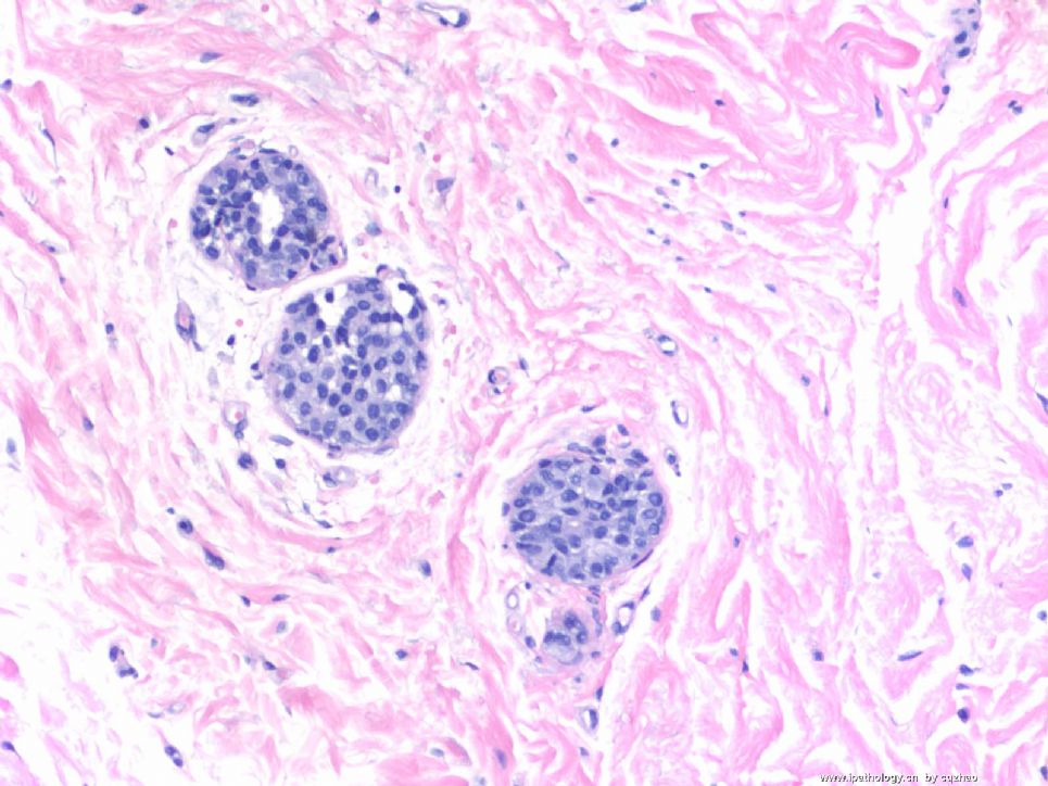
young women (<30 y) had breast reduction. Two sections for each sides were submitted for microscopic examination. See photo for one section. How will you sign out the case?
abin译:年轻女性(<30岁),乳房缩小手术标本。一张切片的两个部位的显微镜下检查。先看第一个部位。你会怎样签发报告?
Fig
200x
名称:图1
描述:图1
-
本帖最后由 于 2008-12-18 20:52:00 编辑
34楼 显示200X视野下三个末梢导管内的增生性病变。对于<30 y的年轻女性,如果没有其它伴随病变,我们在实际工作中可能只是加上一句“上皮生长活跃,建议随访”。
它的性质,我猜可能是小叶增生性病变(小叶瘤变Lobular neoplasia或小叶内瘤变lubular intraepiehtlial neoplasia),相当于LCIS。可能要做E-Ca和p120验证一下。
我不明白为什么间质出现明显的纤维化?

华夏病理/粉蓝医疗
为基层医院病理科提供全面解决方案,
努力让人人享有便捷准确可靠的病理诊断服务。
-
本帖最后由 于 2008-12-23 23:05:00 编辑
For the women younger than 40 with breast reduction we submit 4 slides each side for microscopic examination. This photo shows focal atypical proliferation. We always order IHC for this kind of lesions. Based on morphology it may be a lobular lesion. If they are not lobular lesion, they are normal ducts. In US, there are two different systems for lobular lesions.
1) ALH-LCIS
2) lobular neoplasia 1, 2, 3
Now most people use ALH-LCIS system.
the lesion is very small. It should ALH, not LCIS.
The reasons I put here for this case are
A. For small lesion as above E-cad or/p120 should be performed to confirm the nature of lobular lesion.
B. We should submit more sections if we confirm it is lobular lesion. We generally submit another 10 sections for breast reduction specimen. Remember that ALH or LCIS is a risk factor for cancer.
abin译:
对于40岁以下妇女的乳房缩小手术标本,我们每侧取4张切片作镜下检查。图示局灶性不典型增生。这种病变我们通常要做免疫组化。根据形态学,它可能是小叶病变。如果它们不是小叶病变,就是正常导管。在美国,小叶病变有两种不同的分类系统:
1)ALH-LCIS(不典型小叶增生-小叶原位癌)
2)小叶瘤变 1,2,3级
现在大多数使用ALH-LCIS系统。
病变很小,应该是ALD,而不是LCIS。
我提供这一例的目的:
A、小灶病变应该做E-cad or/p120 以确定小叶病变的性质。
B、如果确定它是小叶病变,应该更多取材。对于乳房缩小标本我们一般再取10块组织。记住ALH或LCIS是进展为癌的危险因素。
我理解的分级:
ALH-LCIS:明确的小叶病变,一个TDLU内一半以上的腺泡被累及(充满且扩张,中央无腔隙),为LCIS。不足一半的腺泡被累及,仅部分充满腺泡,无或仅有轻度扩张,为ALH。LCIS不分级。
LIN 1,2,3级(Bratthauer GL, Tavassoli FA, 2002):1级相当于ALH,2级相当于LCIS,3级:增殖的细胞完全充满并使末梢导管最大限度地扩张到几乎相互融合,或增殖的细胞有明显异型性(多形细胞型),或增殖的细胞完全由印戒细胞组成(印戒细胞型)。多形细胞型和印戒细胞型可出现坏死和钙化,这两型不要求腺泡明显扩张。

华夏病理/粉蓝医疗
为基层医院病理科提供全面解决方案,
努力让人人享有便捷准确可靠的病理诊断服务。
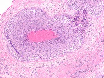
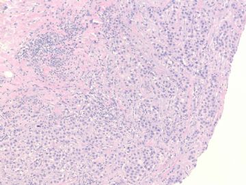
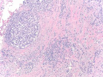
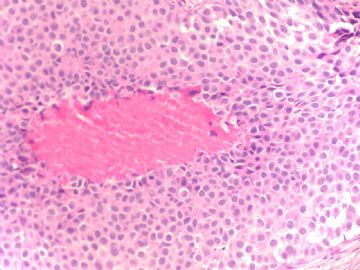


 浸润性导管癌


