| 图片: | |
|---|---|
| 名称: | |
| 描述: | |
- B1301Uterine high grade malignant tumor with divergent differentiation (cqz3)
| 姓 名: | ××× | 性别: | 年龄: | ||
| 标本名称: | |||||
| 简要病史: | |||||
| 肉眼检查: | |||||
Share a case of this week.
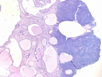
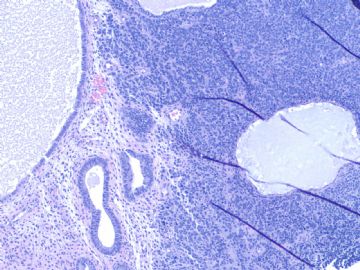
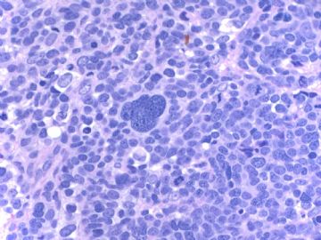
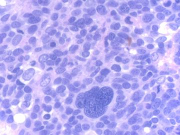
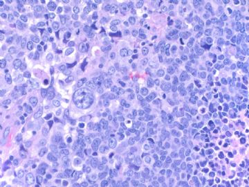
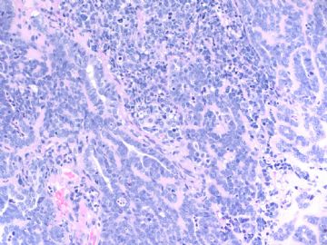
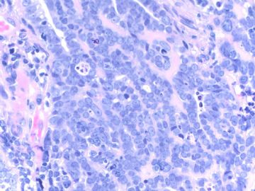
Old lady with atrophic endometrium showing tumor mass in the surface of cystic atrophic endometrium (first figure)
F1 20x
F2 100x
F3 200x
F4-5 400x
F6 200x
F7 400x
F6 and 7 showing focal glandular lesion mixed with other solid lesion.
Your dx or differential dx
-
本帖最后由 于 2009-02-25 09:51:00 编辑
相关帖子
- • 来一例简单罕见的(有诊断)
- • 是肉瘤吗?
- • 子宫内膜,复杂性增生?癌?(有大体结果了)
- • 子宫肿物(透明细胞平滑肌瘤?)
- • 子宫肌壁间肿物
- • 子宫腔内占位。
- • 子宫肿瘤
- • 子宫肌层浸润性癌
- • 8 个子宫上皮肿瘤病例-扫描图片
- • 子宫平滑肌肿瘤?间质肉瘤?
-
stevenshen 离线
- 帖子:343
- 粉蓝豆:2
- 经验:343
- 注册时间:2008-06-03
- 加关注 | 发消息
| 以下是引用cqzhao在2008-12-6 11:26:00的发言:
Most cases I sent here have some difficulties. Hope pathology colleaques what you will do if it is your true case. It is better to mention your differential dx based on H&E. What IHC do you want to order based on your differential dx. In this way you can truely learn sth from studying the case. Thanks |
译文:我提供的大多数病例都是有一些难度。希望病理同仁应该做到这样:即如果该病例是我的,我该怎么做。根据HE切片提出你的鉴别诊断,然后选择哪些免疫标记,只有这样你才能从该病例中学到东西。
非常赞同赵老师的意见。
EIC likes carcinoma in situ, meaning carcinoma limited within glands. EIC is considered as precursor of serous carcinoma in endometrium. Wenxing Zhang, Chinese American pathologist did a lot of research in this topic.
Cytomorphologic features of this case have no any similarity to EIC.
Most cases I sent here have some difficulties. Hope pathology colleaques what you will do if it is your true case. It is better to mention your differential dx based on H&E. What IHC do you want to order based on your differential dx. In this way you can truely learn sth from studying the case.
Thanks
-
stevenshen 离线
- 帖子:343
- 粉蓝豆:2
- 经验:343
- 注册时间:2008-06-03
- 加关注 | 发消息
