


| 图片: | |
|---|---|
| 名称: | |
| 描述: | |
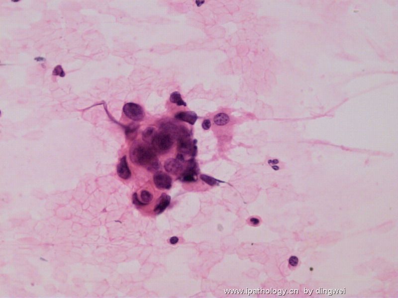
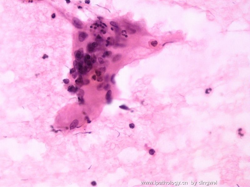
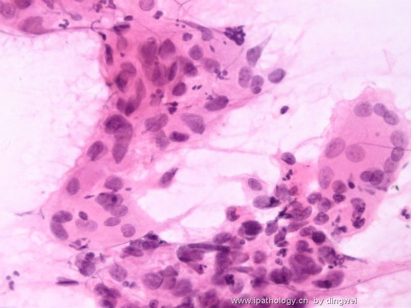
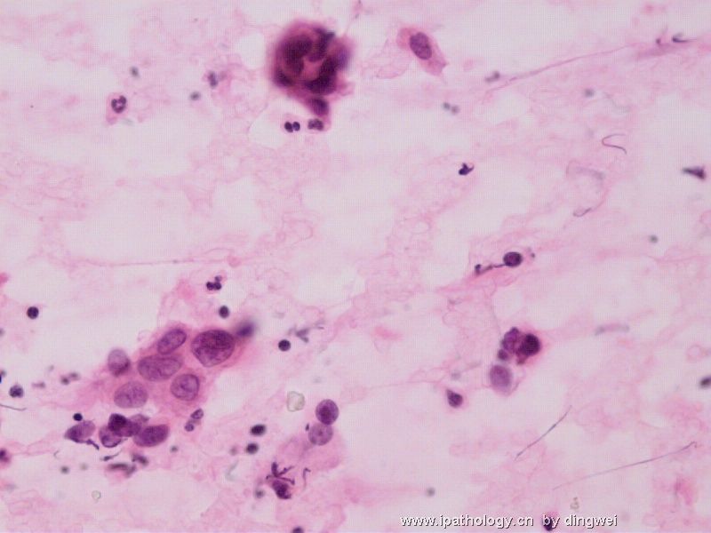
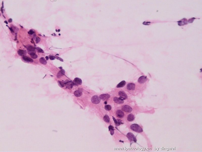
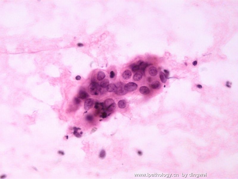
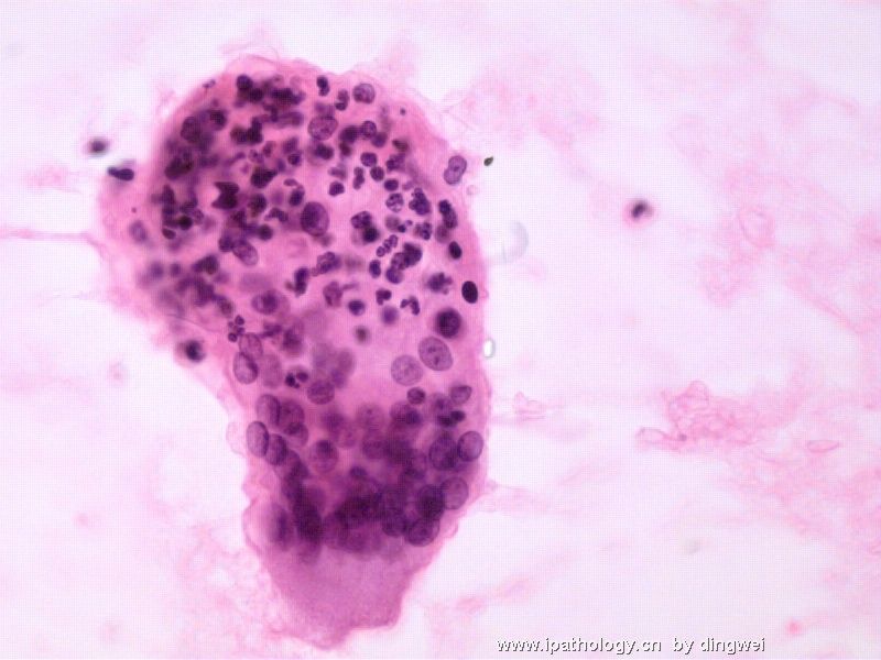
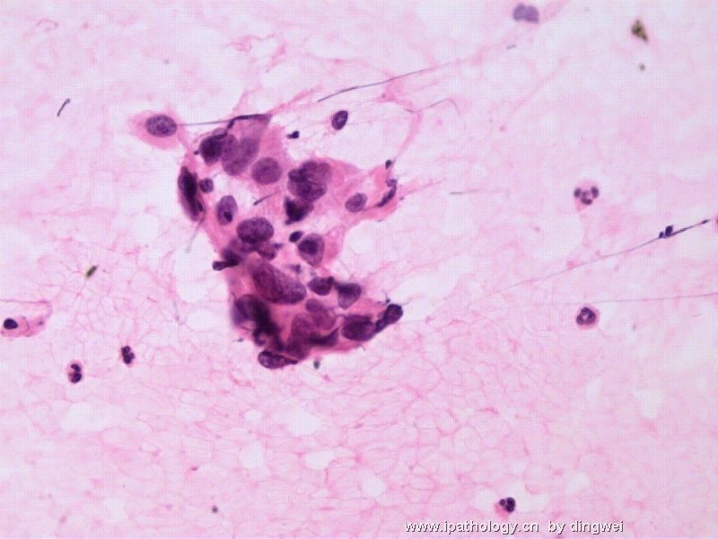
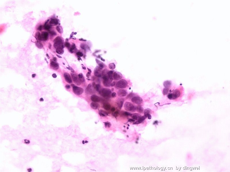
- 锁骨淋巴结穿刺
-
lfl001200546 离线

- 帖子:2808
- 粉蓝豆:40
- 经验:2808
- 注册时间:2007-02-14
- 加关注 | 发消息
Some nuclear features mimic papillary thyroid carcinoma such as nuclear grooves and psudoinclusion. Multinucleated giant cells can also be seen in papillary thyroid carcinoma. But it is unusual to see necrosis in PTC and also unusual in male (can happen but much less often then in female).
Another possibility is metastatic squamous cell carcinoma. Necrotic background is common and multinucleated giant cells can be seen too. One group resembles squamou whorl and one group looks like squamous hugging. But the chromatin is kind of pale and the cytoplasm doesn't show dense squamous differentiation too. Well, there are non-keratinizing SCC where squamous differentiation is hard to see.
I feel the nuclear crowdness and overlappins are really suggestive of a malignant tumor.
Another differential but unlikely consideration is: langerhan's cell histiocytosis: the cells shown all have nuclear gooves and somehow have histiocytic appearance. But i have never seen a case of langerhan's cell histiocytosis with necrosis.
Cannot wait for the answer.
My appology for typing too much english. Thanks Quan Zhi and Abin for the excellent translation in other messages.
| 以下是引用mingfuyu在2008-7-19 10:25:00的发言:
Some nuclear features mimic papillary thyroid carcinoma such as nuclear grooves and psudoinclusion. Multinucleated giant cells can also be seen in papillary thyroid carcinoma. But it is unusual to see necrosis in PTC and also unusual in male (can happen but much less often then in female). Another possibility is metastatic squamous cell carcinoma. Necrotic background is common and multinucleated giant cells can be seen too. One group resembles squamou whorl and one group looks like squamous hugging. But the chromatin is kind of pale and the cytoplasm doesn't show dense squamous differentiation too. Well, there are non-keratinizing SCC where squamous differentiation is hard to see. I feel the nuclear crowdness and overlappins are really suggestive of a malignant tumor. Another differential but unlikely consideration is: langerhan's cell histiocytosis: the cells shown all have nuclear gooves and somehow have histiocytic appearance. But i have never seen a case of langerhan's cell histiocytosis with necrosis. Cannot wait for the answer. My appology for typing too much english. Thanks Quan Zhi and Abin for the excellent translation in other messages. |
您分析的太好了,而且很全面。学习了。
这一例的确是像您说的一样,是食道鳞癌淋巴结转移,由于坏死,组织细胞吞噬,因而出现了大量的多核巨细胞。

- 本人观点纯属个人偏见,希望大家独立判断,正确引用!中华病理技术网:http://www.zhbljs.com/
-
liguoxia71 离线

- 帖子:4174
- 粉蓝豆:3122
- 经验:4677
- 注册时间:2007-04-01
- 加关注 | 发消息


