
 - Paraneoplastic encephalitis and sensory polyneuropathy associated with small cell carcin..图1")
 - Paraneoplastic encephalitis and sensory polyneuropathy associated with small cell carcin..图2")
 - Paraneoplastic encephalitis and sensory polyneuropathy associated with small cell carcin..图3")
 - Paraneoplastic encephalitis and sensory polyneuropathy associated with small cell carcin..图4")
 - Paraneoplastic encephalitis and sensory polyneuropathy associated with small cell carcin..图5")
 - Paraneoplastic encephalitis and sensory polyneuropathy associated with small cell carcin..图6")
 - Paraneoplastic encephalitis and sensory polyneuropathy associated with small cell carcin..图7")
 - Paraneoplastic encephalitis and sensory polyneuropathy associated with small cell carcin..图8")
 - Paraneoplastic encephalitis and sensory polyneuropathy associated with small cell carcin..图9")
 - Paraneoplastic encephalitis and sensory polyneuropathy associated with small cell carcin..图10")
 - Paraneoplastic encephalitis and sensory polyneuropathy associated with small cell carcin..图11")
 - Paraneoplastic encephalitis and sensory polyneuropathy associated with small cell carcin..图12")
 - Paraneoplastic encephalitis and sensory polyneuropathy associated with small cell carcin..图13")
 - Paraneoplastic encephalitis and sensory polyneuropathy associated with small cell carcin..图14")
 - Paraneoplastic encephalitis and sensory polyneuropathy associated with small cell carcin..图15")
 - Paraneoplastic encephalitis and sensory polyneuropathy associated with small cell carcin..图16")

| 图片: | |
|---|---|
| 名称: | |
| 描述: | |
- NP (6) - Paraneoplastic encephalitis and sensory polyneuropathy associated with small cell carcin..
-
These photos are taken from brain, spinal cord, nerve roots, and dorsal root gangia removed from a 59 year-old woman at autopsy. All sections were stained by the luxol fast blue/H&E method. The patient experienced worsening numbness and weakness of lower extremities with difficulty walking for 6 months prior to death due to pneumonia and secondary sepsis. Left sural nerve biopsy and gastrocnemius muscle biopsy prior to death showed severe axonal neuropathy and chronic neurogenic atrophy, respectively. Brain MRI was normal. What is your diagnosis?
 - Paraneoplastic encephalitis and sensory polyneuropathy associated with small cell carcin..图1") 图1
图1 - Paraneoplastic encephalitis and sensory polyneuropathy associated with small cell carcin..图2") 图2
图2 - Paraneoplastic encephalitis and sensory polyneuropathy associated with small cell carcin..图3") 图3
图3 - Paraneoplastic encephalitis and sensory polyneuropathy associated with small cell carcin..图4") 图4
图4 - Paraneoplastic encephalitis and sensory polyneuropathy associated with small cell carcin..图5") 图5
图5 - Paraneoplastic encephalitis and sensory polyneuropathy associated with small cell carcin..图6") 图6
图6 - Paraneoplastic encephalitis and sensory polyneuropathy associated with small cell carcin..图7") 图7
图7 - Paraneoplastic encephalitis and sensory polyneuropathy associated with small cell carcin..图8") 图8
图8 - Paraneoplastic encephalitis and sensory polyneuropathy associated with small cell carcin..图9") 图9
图9 - Paraneoplastic encephalitis and sensory polyneuropathy associated with small cell carcin..图10") 图10
图10 - Paraneoplastic encephalitis and sensory polyneuropathy associated with small cell carcin..图11") 图11
图11 - Paraneoplastic encephalitis and sensory polyneuropathy associated with small cell carcin..图12") 图12
图12 - Paraneoplastic encephalitis and sensory polyneuropathy associated with small cell carcin..图13") 图13
图13 - Paraneoplastic encephalitis and sensory polyneuropathy associated with small cell carcin..图14") 图14
图14 - Paraneoplastic encephalitis and sensory polyneuropathy associated with small cell carcin..图15") 图15
图15 - Paraneoplastic encephalitis and sensory polyneuropathy associated with small cell carcin..图16") 图16
图16
1. Left frontal lobe neocortex
2-3. Left transentorhinal cortex
4. Substantia nigra
5. Nucleus of oculomotor nerve (midbrain)
6. Cerebellar cortex
7-8. Cerebellar dentate nucleus
9. Basis pontis
10-11. Inferior olivary nucleus of medulla oblongata
12. Lumbar spinal cord
13. Anterior nerve root (lumbar)
14. Posterior nerve root (lumbar)
15-16. Lumbar dorsal root ganglion
标签:
-
本帖最后由 于 2007-01-22 11:31:00 编辑

聞道有先後,術業有專攻
×参考诊断
Paraneoplastic syndromes are diseases caused indirectly by the presence of malignancy. They are the results of hormones or cytokines elaborated by cancer cells, or of the body's immune response to cancer cells. Sometimes, paraneoplastic syndromes are the first signs of cancer (i.e., before the cancer is diagnosed). The types of cancer involved are not disease-specific. Lung (both small cell and non-small cell types), ovarian and breast cancers are the common offenders, but cancers of many other organs have been reported.
There are four categories of paraneoplastic syndromes - hematological, neurological, mucocutaneous, and endocrine/metabolic. Neurological paraneoplastic syndromes may affect the central and/or the peripheral nervous systems. Many neurological paraneoplastic syndromes or disorders are caused by autoimmune mechanisms, with cancer-induced circulating autoantibodies directed against functional components at various parts of our nervous system. These functional elements include neurons and proteins on cellular surface. Depending on their locations and functions, different neurological disorders arise. Some examples are listed below.
1. Limbic system (hippocampus/medial temporal cortex) - seizures, acute or subacute dementia (limbic encephalitis)
2. Brainstem - cranial nerve palsy, seizures, sudden death (brainstem encephalitis)
3. Cerebellar Purkinje cells - ataxia (cerebellitis)
4. Spinal cord - pain and weakness (myelitis)
5. Posterior root ganglia - pain and numbness (ganglionitis and sensory neuropathy)
6. Myelin sheaths of nerve fibers - numbness and weakness (demyelinating sensorimotor polyneuropathy)
7. Calcium channel at neuromuscular junction - muscle weakness (Lambert-Eaton myasthenic syndrome)
8. Voltage-gated potassium channel on neurons - persistent muscular contraction (neuromyotonia), excessive sweating, sleep disorder, memory loss, hallucinations and delusions
9. Endothelial cells in skeletal muscle and dermis - skin rashes and muscle weakness with inflammation (dermatomyositis)
From the list above, it is not difficult to realize that manifestations of paraneoplastic neurological disorders vary from case to case and often mislead clinicians. Since circulating autoantibodies are often present in the serum of these patients, screening for them may occasionally be diagnostic. However, only a portion of targeted cells and proteins has been characterized so screening results are often false negative. Biopsy of tissue before death is often non-diagnostic since pathology is non-specific and often very focal. Often the final diagnosis is rendered by a complete autopsy. Fortunately, empiric (but expensive) therapy with plasmapheresis and/or IVIg may result in dramatic alleviation of the symptoms and, if implemented early, may even promise partial functional recovery. A high index of clinical suspicion, thorough oncologic workup, and complete resection, effective radiation therapy or chemotherapy of the cancer found often is curative for the associated paraneoplastic syndrome.
聞道有先後,術業有專攻
-
本帖最后由 于 2007-01-22 11:32:00 编辑
This is a rare case of paraneoplastic (autoimmune) encephalitis and sensory polyneuropathy caused by undiagnosed metastatic small cell carcinoma in the mediastinum, most probably of pulmonary origin.
Photos uploaded in the first batch reflect the extent of inflammation in the central nervous system, and degeneration of the spinal cord and the peripheral nervous system. Figures 1 and 6 show normal frontal lobe isocortex and normal cerebellar cortex (including intact Purkinje cells that often are targets of paraneoplastic or autoimmune encephalomyelitis) without inflammation. Figures 2-3 show perivascular inflammation and microglial nodules in transentorhinal cortex (medical temporal lobe) indicative of active mesial temporal encephalitis. Figures 4 and 6-11 show varying numbers of microglial nodules with or without neuronophagia in varying parts of the brainstem and cerebellum, indicative of active brainstem encephalitis and cerebellitis. Figures 12 shows degeneration (loss of blue myelin staining) of the posterior column of the spinal cord due to severe sensory polyneuropathy. Figure 13 shows relatively normal anterior spinal nerve root (motor), and Figure 14 shows the markedly degenerated posterior spinal nerve root (sensory). Note the abundant myelinated nerve fibers in the former and relatively few remaining myelinated nerve fibers in the latter.
Figures 15 and 16 show many small lymphocyte-like cells in the posterior root ganglia, suggestive of active ganglionitis, but immunohistochemical stain (not shown) shows these cells to be negative for CD45. Therefore, they probably represent satellite cells normally present around ganglion cells in dorsal root ganglia.
The four photos uploaded in the second batch are diagnostic of small cell carcinoma, most probably of pulmonary origin.
聞道有先後,術業有專攻
-
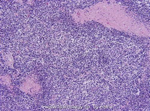
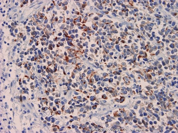
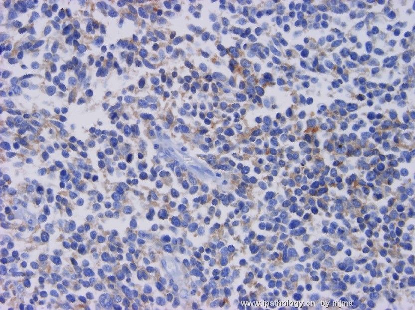
Sorry for this delayed follow-up. At the autopsy, a 2 cm enlarged mediastinal lymph node was found. No tumor was identified in both lungs or other organs. Figures 1 and 2 are H&E stained section of the enlarged mediastinal lymph node. Figure 3 is Cam5.2 immunostain, and figure 4 is synaptophysin immunostain. With these additional findings, how would you explain the entire case?
 图1
图1 图2
图2 图3
图3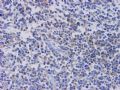 图4
图4
聞道有先後,術業有專攻


