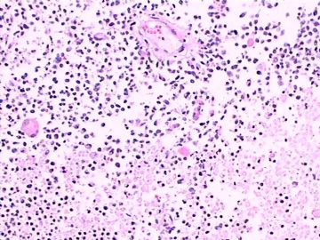
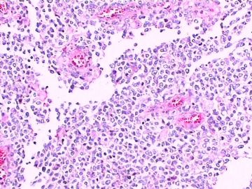
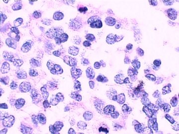
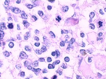
| 图片: | |
|---|---|
| 名称: | |
| 描述: | |
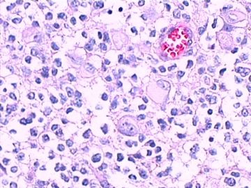
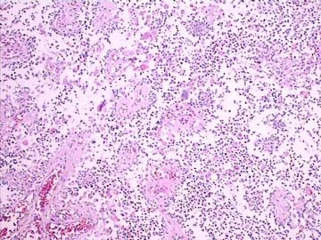
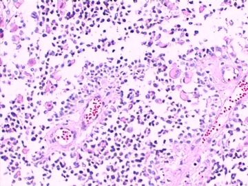
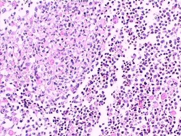
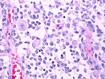
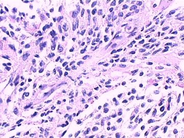
- 左额叶脑肿块(诊断?)请各位老师发表高见。
| 姓 名: | ××× | 性别: | 女 | 年龄: | 70岁 |
| 标本名称: | 左额叶病变组织 | ||||
| 简要病史: | 头部不适伴语言迟缓7天入院。 | ||||
| 肉眼检查: | 肿瘤组织3块,大小共4.2x2.2x0.6cm,切面灰红,质软。 | ||||
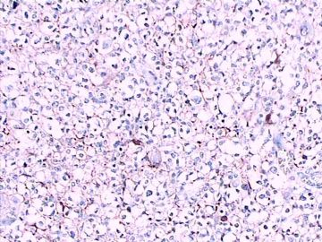
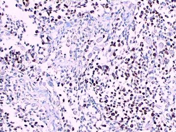
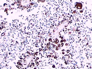
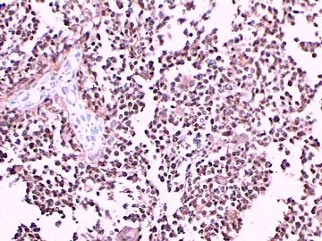
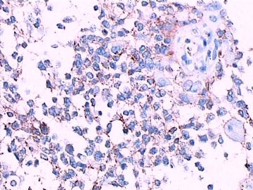
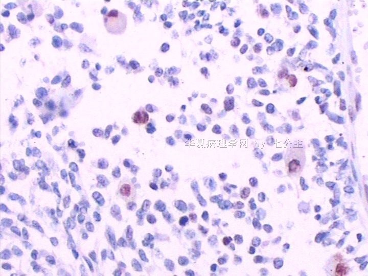
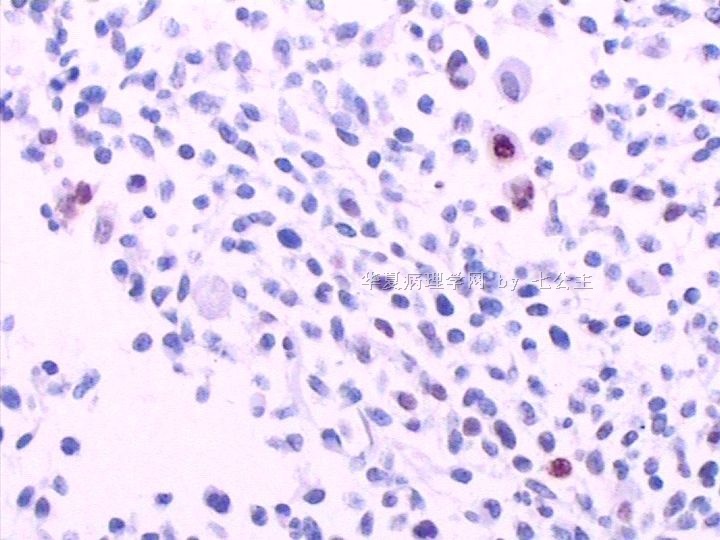
免疫组化染色:GFAP胞浆丰富的大细胞阳性,Olig2小细胞阳性,Syn阳性,S-100阳性,MBP部分细胞阳性,Neun局部散在少量大细胞阳性,需要除外残余神经元;Ki-67阳性率约40%。
11图MBP,12图Olig2,13图GFAP,14图S-100,15图Syn。
-
本帖最后由 于 2011-01-02 13:23:00 编辑
At this age with tumor necrosis, 1st choice should be GBM, sometimes, more immunostaining just making you more confusion,
Like this oligo2 marker, some paper mentioned it is a stem cell marker, due to its non-specific, not widely use yet, that may be
the reason it is called oligo 2 but not oligo 1. Remember, any glial tumor may have intrapped neurons and oligo cells in the tumor, you do more
staining, making you harder to make a dx.
good good study, day day up.
happy new horse year.
-
本帖最后由 于 2011-01-03 19:54:00 编辑
1)本病例有坏死、明显的核分裂象、Ki-67 指数40%,因此定位于WHO3级的脑肿瘤比较合理。
2)组织学分类:根据形态和IHC标记基本特点,主要鉴别为少突胶质细胞伴神经细胞分化和节细胞胶质瘤;
3)少突胶质瘤中出现神经细胞有二种情况,1)肿瘤中残留的正常胶质细胞;2)肿瘤伴有神经细胞分化(oligodendroglioma with neurocytic differentiation)。
4)鉴别要点是肿瘤中神经细胞的数量、分布和形态。
5)本病例有坏死、核分裂多;Olig2+、Syn+、GFAP+、 Ki-67指数高,比较符合“间变性少突胶质细胞伴神经细胞分化, WHO3级)。
仅供参考。
谢谢!

- xljin8
| 以下是引用海上明月在2011-1-1 23:14:00的发言: 这不是少枝(突)胶质瘤,但细胞形态和组织结构有少枝胶质瘤样的表现。主要形态:(1)小血管增生比较明显,可见血管周假菊形团或假乳头状结构。(2)绝大多数瘤细胞为形态较一致的体积较小的细胞,易与少枝胶质瘤混淆。(3)这些小圆细胞胞核呈圆形或卵圆形,核染色质细密分布,少数瘤细胞可见核仁。这些细胞中意见核分裂。(4)一些视野下,可见神经节细胞样细胞,胞体大,胞浆丰富并嗜酸性着色,核院或卵圆形,核仁明显。(5)少数视野下(图10)显示瘤细胞呈长梭形。(6)IHC标记突出显示节细胞样细胞和小细胞均为Syn和S-100阳性,GFAP主要为大细胞阳性,Olig2主要为小细胞阳性,MBP有阳性,Neun局灶大细胞阳性。没有染CgA和CK。 对:一些视野下,可见神经节细胞样细胞,胞体大,胞浆丰富并嗜酸性着色,核院或卵圆形,核仁明显。但这些大细胞GFAP阳性;NeuN阴性呀。大细胞和小细胞均为Syn和S-100阳性。是否是间变型星形细胞瘤? |
| 以下是引用yinwang在2011-1-12 12:39:00的发言: 你提的问题非常好!我想这也是神经病理的难点之处。 SYN阳性,关键你要看它在什么部位,肿瘤细胞胞浆或突起内?还是在残存的神经元周围或神经毡样基质内?该病例肿瘤范围广,累及大脑皮层,因此SYN阳性并不奇怪,这也是胶质瘤弥漫性浸润,无边界的特点。如有可能请追加做一下p53蛋白。鄙人姓汪,名寅。不是王寅。期待p53 蛋白结果。谢谢! 嘿嘿,是我不好,打字过快,未经审阅就发帖子了,重大失误。特向汪寅教授表示深深歉意! 汪寅教授是我国著名的年轻有为临床神经病理学家。谢谢汪教授为我们解惑和指导! |

- 王军臣
| 以下是引用七公主在2011-1-12 18:12:00的发言: 不好意思,将汪老师名字打错了,已改正。不知P53蛋白在胶质母中的意义? 请见以下文献: Brain Tumor Pathol. 2010 Oct;27(2):95-101. Epub 2010 Nov 3. p53 abnormality and tumor invasion in patients with malignant astrocytoma.Momota H, Narita Y, Matsushita Y, Miyakita Y, Shibui S. Neurosurgery Division, National Cancer Center Hospital, 5-1-1, Tsukiji, Chuo-ku, Tokyo, 104-0045, Japan. momota-nsu@umin.ac.jp AbstractMalignant astrocytomas are characterized by diffusely infiltrating nature, and the abnormality of p53 is a cytogenetic hallmark of astrocytic tumors. To elucidate the relationship between p53 abnormality and invasiveness of the tumors, we studied mutation and protein expression of p53 in 48 consecutive patients with malignant astrocytoma (14 anaplastic astrocytomas and 34 glioblastoma multiformes). The tumors were classified into three categories according to the features of magnetic resonance imaging, and 5, 7, and 36 tumors were classified into diffuse, multiple, and single type, respectively. We then examined how these tumor types correlate with MIB-1 staining index, TP53 gene mutation, and p53 protein expression. We found that p53 immunopositivity or TP53 mutation was frequently observed in diffuse and multiple types. These abnormalities of p53 were also associated with high MIB-1 staining index and strong expression of vascular endothelial growth factor. Furthermore, diffuse- and multiple-type tumors were significantly correlated with poor progression-free survival, whereas only multiple-type tumors were significantly correlated with poor overall survival. As diffuse and multiple features on imaging modalities represent invasive characteristics of the tumors, p53 abnormalities may affect the invasive and aggressive nature of malignant astrocytomas. |

- 王军臣
