| 图片: | |
|---|---|
| 名称: | |
| 描述: | |
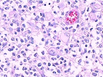
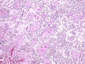
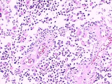
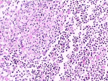
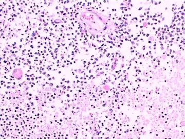
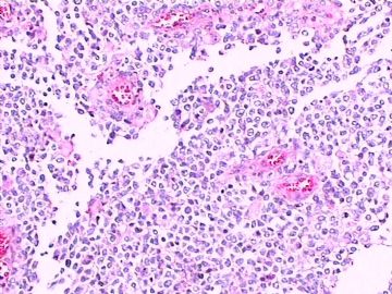
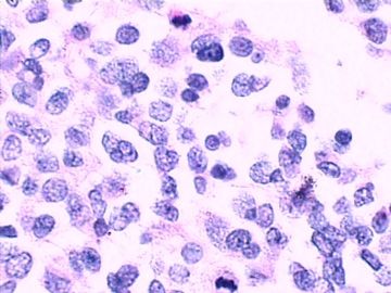
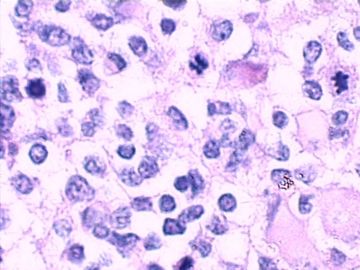
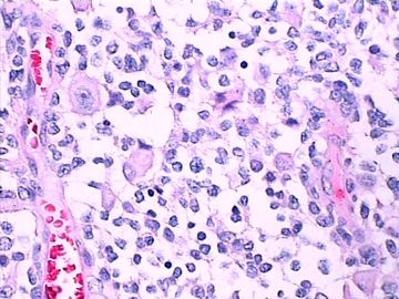
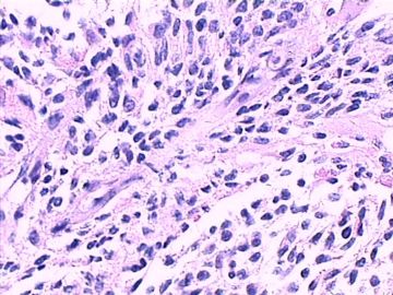
- 左额叶脑肿块(诊断?)请各位老师发表高见。
| 姓 名: | ××× | 性别: | 女 | 年龄: | 70岁 |
| 标本名称: | 左额叶病变组织 | ||||
| 简要病史: | 头部不适伴语言迟缓7天入院。 | ||||
| 肉眼检查: | 肿瘤组织3块,大小共4.2x2.2x0.6cm,切面灰红,质软。 | ||||
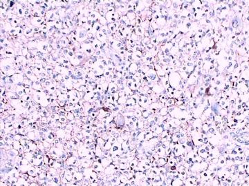
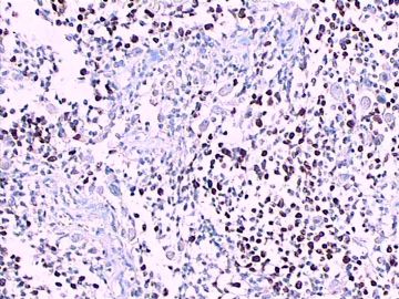
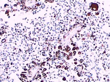
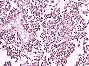
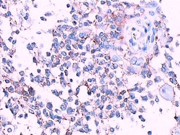
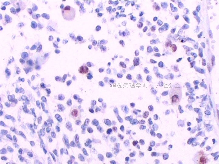
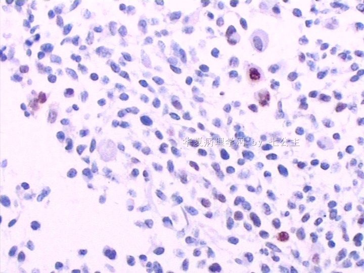
免疫组化染色:GFAP胞浆丰富的大细胞阳性,Olig2小细胞阳性,Syn阳性,S-100阳性,MBP部分细胞阳性,Neun局部散在少量大细胞阳性,需要除外残余神经元;Ki-67阳性率约40%。
11图MBP,12图Olig2,13图GFAP,14图S-100,15图Syn。
标签:
-
本帖最后由 于 2011-01-02 13:23:00 编辑
×参考诊断
多形性胶质母细胞瘤,WHO4级
At this age with tumor necrosis, 1st choice should be GBM, sometimes, more immunostaining just making you more confusion,
Like this oligo2 marker, some paper mentioned it is a stem cell marker, due to its non-specific, not widely use yet, that may be
the reason it is called oligo 2 but not oligo 1. Remember, any glial tumor may have intrapped neurons and oligo cells in the tumor, you do more
staining, making you harder to make a dx.
good good study, day day up.
happy new horse year.
-
benben520sps 离线
- 帖子:1045
- 粉蓝豆:568
- 经验:1254
- 注册时间:2009-07-28
- 加关注 | 发消息
-
benben520sps 离线
- 帖子:1045
- 粉蓝豆:568
- 经验:1254
- 注册时间:2009-07-28
- 加关注 | 发消息
-
jianshu322 离线
- 帖子:447
- 粉蓝豆:17
- 经验:1405
- 注册时间:2008-12-22
- 加关注 | 发消息
