| 图片: | |
|---|---|
| 名称: | |
| 描述: | |
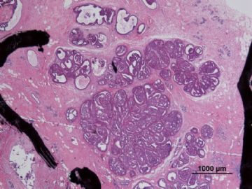
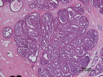
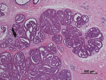
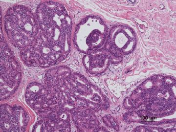
- B3087有难度的乳头状肿瘤病例(非典型性周围型导管内乳头状瘤还是周围型导管内乳头状瘤伴DCIS?)新加病灶测..
| 姓 名: | ××× | 性别: | F | 年龄: | 37 |
| 标本名称: | |||||
| 简要病史: | |||||
| 肉眼检查: | |||||
病历摘要:
“发现左乳肿物1年余,右乳肿物1周”入院。
2010年12月6日双乳钼靶片示:双乳呈混合型Ⅳb(纤维囊性增生为主),左乳外上区多发肿块不除外恶变可能,建议手术,右乳外上区良性增生结节,BI-RADS:III,左乳BI-RADS:V;
查乳腺彩超示:符合双侧乳腺增生声像,左乳头外上象限低回声团块,考虑为实性占位,不除外恶变可能,建议病理检查,右乳低回声肿块,考虑纤维腺瘤可能。
专科检查:双乳外形欠对称,左乳12-2点位距乳头2cm可触及一肿物,大小约4.0×5.0cm,质硬,边界欠清,表面欠光滑,活动度欠佳,与皮肤、胸壁无粘连。右乳11点位距乳头2cm可触及一肿物,大小约1.5×1.5cm,质韧,边界尚清,表面尚光滑,活动度一般,与皮肤、胸壁无粘连。
临床诊断:
1.乳房肿物(左乳癌?)2.乳房肿物(右乳增生结节?)3.乳腺增生(双侧)
大体病理检查:
送检(左侧)乳腺肿物切除标本:6.0cm×4.0cm×1.0cm,切面见大小为1.5cm×1.3cm灰红结节。
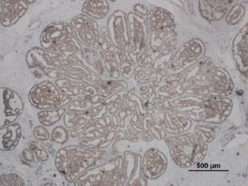
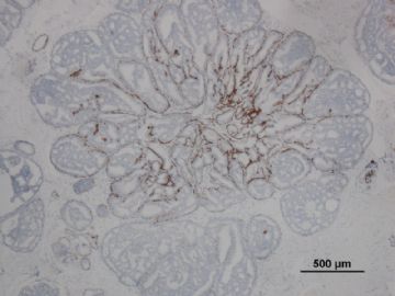
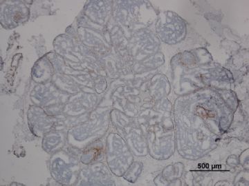
其中一张切片,免疫组化顺序为:ER,CK5/6,CD10
-
本帖最后由 于 2011-01-07 21:28:00 编辑
相关帖子
- • 乳腺包块
- • 左乳癌标本乳头一个导管内的病变
- • 乳腺两个相邻导管内的病变
- • 乳腺肿物
- • 乳腺肿物,请各位老师帮忙会诊
- • 女 46岁发现左乳腺肿块一月余
- • 乳腺包块。33岁
- • 左乳肿块,协助诊断
- • 乳腺肿物
- • 乳腺肿物
| 以下是引用yeqing在2011-1-3 22:30:00的发言:
学习了,谢谢各位老师的分析。 局灶性DICS和ADH形态学上区别的这个度不好掌握啊。 |
It is true it is not easy to differentiate low grade dcis from adh.
Low nuclear grade, small size. This is why i favor ADH above.
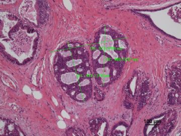
Louzhu, please let us know the size of the focus above.
| 以下是引用cqzhao在2011-1-2 23:39:00的发言:
Pathology diagnosis should have criteria. If you think it is papilloma with DCIS, you have to mention the reasons. If you have good myoepithelial stains, this case should be easy. For this case most important stains are myoepithelial stains. I only saw calponin stain above. |
赵老师意见:
病理诊断要依标准。楼主要想诊断为乳头状瘤伴有DCIS,那就应说出诊断的理由。只要肌上皮标记做得好,本例的诊断就便当了。
本例最要紧的标记是肌上皮标记。可前面只见calponin标记。
注:至少要做核标记物P63.

- 王军臣
-
本帖最后由 于 2011-01-03 12:43:00 编辑
| 以下是引用老山羊在2011-1-1 23:10:00的发言: 我们科内讨论的意见也是集中于是“周围型导管内乳头状瘤伴ADH”还是“周围型导管内乳头状瘤伴DCIS”,最终未达成一致。 |
要说好办法,那就是要选择比较公认的肌上皮标志物做IHC检测,以对肌上皮存在的情况进行评价。一般应选择一个胞核表达、两个浆表达的标志物。赵澄泉老师曾反复强调过这些。
显示肌上皮胞核的最好标志物是:P63
显示胞浆的常用标志物SMA和calponin。也有选择actin,意义差不多。
乳腺肌上皮可具有多种免疫表型,楼主做了CK5/6和CD10。一般来说,CK5/6适于做基底细胞标记,当然也有说做肌上皮的。楼主IHC标记CK5/6显示部分阳性,倒不如做P63再看看。
P63是观察肌上皮存在的一个很好的核型标志物,它避免了SMA、calponin或actin在导管周围的乳腺肌纤维母细胞也会阳性的缺陷。
我们曾用D2-40做为标记检测,显示肌上皮的效果也很不错。
楼主做CD10染色意在显示肌上皮,图片显示其在导管周围阳性表达很连续(不是一般的连续)。
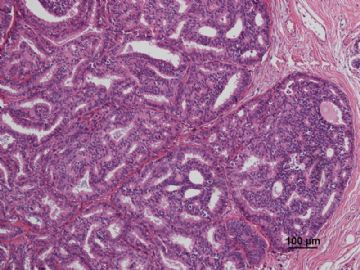
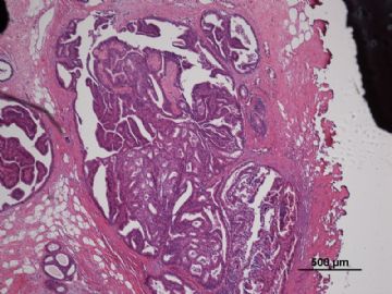
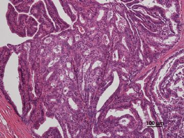
仅从形态学看导管形态和其中窗孔分布形式(本例有的导管内甚至可见“边窗”)及其规则性,加上现有的标记,是不符合DCIS的,但符合非典型乳头状瘤或ADH。
此外,只要标记和形态上表现为导管内增生的细胞有肌上皮的存在,不管有无坏死和细胞异型,可能是不能评判为DCIS的。坏死和细胞异型不是区分ADH和DCIS的鉴别指标,但可以是区分ADH或DCIS究竟是高级别还是低级别的主要指标。
以上仅供参考,不妥之处请指正。

- 王军臣
-
本帖最后由 于 2011-01-03 08:13:00 编辑
非常好的病例和讨论。对诊断为乳头状肿瘤大家无很大分歧,争论的焦点在于是否存在“原位癌”。实际上它涉及到三方面的问题:
一、病理学的诊断问题:
1)乳头状瘤与不典型乳头状瘤的鉴别诊断;
2)不典型乳头状瘤与乳头状原位癌的鉴别诊断;
3)不典型乳头状瘤与DCIS累及乳头状瘤的鉴别;
困难在3,因为不像1和2可依靠肌上皮细胞的存在与否和消失程度做鉴别点。
如果举一个极端的例子-“导管原位癌累及不典型乳头状瘤”,这时就不能单单依靠肌上皮作为唯一的鉴别要点。
二、鉴别诊断的临床意义?
三、当病理诊断有分歧时, 如何打病理报告?
谢谢!

- xljin8
| 以下是引用cqzhao在2011-1-2 23:56:00的发言:
My comment for your department diagnosis 1. 周围型导管内乳头状瘤伴ADH,综合科内讨论意见,未能除外局灶DCIS Based on detaled study or evaluation, if pathologists think it is 周围型导管内乳头状瘤伴ADH, then just call it. Do not need to 未能除外局灶DCIS. If the patient still has DCIS or invasion cancer in her breast, it is ok. We make our diagnosis based on available sample. If you think it is focal DCIS, then just call it. 2. 周围型导管内乳头状瘤伴ADH,综合科内讨论意见,未能除外局灶DCIS is a very conseravtive pathology diagnosis. However it will cause confusion to patients and clinicians 3. As pathologists we should provide correct diagnosis and also know what our clinicians would do based on our diagnosis. We can provide some suggestion for treatment. However clinicians (surgeons or oncologists) will take the responsibility for the treatment plans. 4. Should have more myoepithelial stains, such as p63 or SMMHc if you are not sure the dx 5. Why stain CD10? for your reference Thank 老山羊for sharing the case with us.
|
赵老师对贵科的诊断发表如下意见(中文大意):
1.关于“周围型导管内乳头状瘤伴ADH,综合科内讨论意见,未能除外局灶DCIS”
基于详尽的研究或评价, 一旦病理医生认为是“周围型导管内乳头状瘤伴ADH”, 那就这样诊断好了,无需加上“未能除外局灶DCIS”。要是病人乳腺还患有DCIS或是浸润癌,那也就照样诊断好了。我们下诊断是根据可供诊断的标本来评价的,如果贵方认为可诊断局灶DCIS,那就不要“未能除外”,就报局灶DCIS。
2. 再说关于“周围型导管内乳头状瘤伴ADH,综合科内讨论意见,未能除外局灶DCIS”
这是一个很保守的病理诊断。而这将造成病人和临床医生慌乱。
3. 作为病理医生,我们要提供正确的病理诊断,也要知晓临床医生根据我们的诊断将会做什么样的治疗。我们可以给临床提些治疗建议。而这样,临床医生(不管是外科医生还是肿瘤科医生)做治疗方案时就将其纳入参考。
4. 如果贵方不能确诊,建议再做些肌上皮染色,例如p63和SMMHc.
5. 请问:为啥做CD10标记?
仅供参考!
谢谢Dr.老山羊给我们分享这个病例!

- 王军臣
| 以下是引用老山羊在2011-1-1 23:10:00的发言: 我们科内讨论的意见也是集中于是“周围型导管内乳头状瘤伴ADH”还是“周围型导管内乳头状瘤伴DCIS”,最终未达成一致。 |
My comment for your department diagnosis
1. 周围型导管内乳头状瘤伴ADH,综合科内讨论意见,未能除外局灶DCIS
Based on detaled study or evaluation, if pathologists think it is 周围型导管内乳头状瘤伴ADH, then just call it. Do not need to 未能除外局灶DCIS. If the patient still has DCIS or invasion cancer in her breast, it is ok. We make our diagnosis based on available sample. If you think it is focal DCIS, then just call it.
2. 周围型导管内乳头状瘤伴ADH,综合科内讨论意见,未能除外局灶DCIS is a very conseravtive pathology diagnosis. However it will cause confusion to patients and clinicians
3. As pathologists we should provide correct diagnosis and also know what our clinicians would do based on our diagnosis. We can provide some suggestion for treatment. However clinicians (surgeons or oncologists) will take the responsibility for the treatment plans.
4. Should have more myoepithelial stains, such as p63 or SMMHc if you are not sure the dx
5. Why stain CD10?
for your reference
Thank 老山羊for sharing the case with us.
Pathology diagnosis should have criteria. If you think it is papilloma with DCIS, you have to mention the reasons. If you have good myoepithelial stains, this case should be easy.
For this case most important stains are myoepithelial stains. I only saw calponin stain above.
-
本帖最后由 于 2011-01-01 23:13:00 编辑
我们科内讨论的意见也是集中于是“周围型导管内乳头状瘤伴ADH”还是“周围型导管内乳头状瘤伴DCIS”,最终未达成一致。
最后病理报告:“周围型导管内乳头状瘤伴ADH,综合科内讨论意见,未能除外局灶DCIS”。病灶范围较广,临床医生综合术中情况可能未完全切除病灶,与患者及家属充分讨论后行第二次手术(乳腺皮下腺体完全切除),病理:“见小灶不典型导管内乳头状瘤,切缘阴性。建议临床密切随访”。
第1楼内的病灶,如果理解为乳头状瘤,则考虑为乳头状瘤伴ADH,但仔细观察,每个增生的导管相互分离,未见纤维血管轴心,是否可理解为不典型增生导管的集合?结合病灶大小,是否够DCIS标准?我个人是比较倾向于“周围型导管内乳头状瘤伴DCIS”的。
实际工作中,总会有一些类似的交界性病变,让我们左右为难。
也希望大家说一说处理类似问题的好办法!

