| 图片: | |
|---|---|
| 名称: | |
| 描述: | |
- B2945Clinical and biological significance of E-cadherin protein expression in invasive lobular carcinom..
| 姓 名: | ××× | 性别: | 年龄: | ||
| 标本名称: | |||||
| 简要病史: | |||||
| 肉眼检查: | |||||
Am J Surg Pathol. 2010 Oct;34(10):1472-9.
Clinical and biological significance of E-cadherin protein expression in invasive lobular carcinoma of the breast.
Rakha EA, Patel A, Powe DG, Benhasouna A, Green AR, Lambros MB, Reis-Filho JS, Ellis IO.
Department of Histopathology, Nottingham University Hospitals NHS Trust, UK. emadrakha@yahoo.com
Abstract
BACKGROUND: Although virtually all cases of lobular carcinoma in situ lack E-cadherin expression, a proportion of morphologically typical invasive lobular carcinomas (ILCs) retain its expression. The frequency and significance of E-cadherin expression in ILC remain to be elucidated. In this study, we have assessed E-cadherin protein expression in a well-characterized series of histologically defined ILC (239 cases) with a long-term clinical follow-up to determine the frequency, clinical and biological significance of its expression. E-cadherin-positive ILCs (ILC+) were subsequently examined to assess the expression of component members of the E-cadherin membrane complex (E-cadherin, p120, α, β, and γ-catenins) to determine its integrity.
RESULTS: Thirty-eight ILC cases (16%) showed positive E-cadherin expression (ILC+). Membranous expression of E-cadherin was mainly circumferential with frequent coexisting perimembranous cytoplasmic expression. No association between E-cadherin expression and any of the clinicopathologic variables, immunophenotype, or tumor behavior was identified, apart from an association with lobular histologic subtype and vascular invasion. Analysis of the E-cadherin-catenin complex showed abnormal expression of one or more of the catenin complex members in the majority of cases. The most frequent observation was the diffuse cytoplasmic expression of catenins, in particular p120, which showed similar expression to that reported in E-cadherin-negative ILCs.
CONCLUSIONS: These results provide evidence that E-cadherin is expressed in a proportion of ILC, however, unlike ductal carcinoma, its expression seems to be of limited significance and it is usually associated with evidence of impaired integrity of the E-cadherin-catenin membrane complex. Our data offer a possible explanation for the presence but lack functionality of E-cadherin in some cases of ILC and imply that immunohistochemical expression of E-cadherin per se in ILC histologic phenotypic tumors should not preclude its diagnosis.
PMID: 20871222 [PubMed - indexed for MEDLINE]
-
本帖最后由 于 2010-11-28 08:16:00 编辑
相关帖子
-
本帖最后由 于 2010-11-28 17:49:00 编辑
非常有意义的研究报道,对传统上用“癌细胞膜表达E- cadherin” 做为鉴别IDC和ILC的标记提出了挑战。
形态学典型的ILC16% 可细胞膜表达E-cadherin, 但是P-120仍为细胞质表达(ILC型)。
因此,具有经典形态的浸润性小叶癌E-cadherin 和P120 的表道方式可以归纳为。
1)Ecadherin 细胞膜不表达(或明显弱表达);P120细胞质表达,
2)E-cadherin 膜表达;P120 细胞质表达。
第二情况的发生率可高达16%, 因此,我们应该注意到形态学典型的浸润小叶癌可以细胞膜表达E-cadherin, 此时标记P120 有利于鉴别。但是其临床预后与E-cadherin-的ILC无明显区别(除浸润血管外)。作者认为E-cad+ILC中E-cadherin是无功能性的。
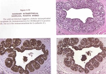
此外,要注意少数乳腺导管上皮内肿瘤,单纯根据HE形态无法区别是DCIS还是LCIS(见图)。 用IHC标记E-cadherin 和34BE12可有4种表达形式:
1)E-cadherin+34BE12-(DCIS表型)
2)E-cadherin-34BE12+(LCIS表型)
3)E-cadnerin-s4BE12-(阴性杂交)
4) E-cadherin+34BE12+(阳性杂交)
第3和第4种免疫表型被认为是Mammary intraepithelial neoplasia MIN) 。
Dr. Tavasoli推测MIN可能为乳腺干细胞性的病变。MIN可能反应乳腺干细胞肿瘤或最早期后干细胞。它具有塑形和演变成导管内或小叶内上皮肿瘤和浸润性乳腺癌的潜能。MIN临床处理,注释,分级、处理根据免疫表型。目前认为:E-cadherin是导管上皮病变特征E-cadherin + 按DIN处理;E-cadherin - 按LIN处理
(参考文献:AFIP Tumors of the breast, series 4Page 94-95.)
DR. Zhao 提供了非常好的文章,它纯清了我们在日常工作中的疑问“形态学典型的浸润性小叶, IHC 却细胞膜表达E-cadherin,并提出对于这种情况标记P120 可帮助鉴别。
由于未看全文,可能理解有误,请Dr.Zhao 指正,谢谢!

- xljin8
| 以下是引用cqzhao在2010-11-11 4:30:00的发言:
Above photos-E-cadherin: Authors interpretate D as invasive ductal ca only. Allother photos are invasive lobular ca. Do you agree?
|
Authors interpretate F IDC, others ILC ? NOT D as IDC?
我觉得F是浸润性导管癌,CD是浸润性小叶癌,A看不清细胞特点不好判断,B如果是浸润性小叶癌则是多形性亚型,D可能不是浸润性小叶癌。
染色结果不容易理解。可能需要结合原切片的阳性、阴性对照考虑。

华夏病理/粉蓝医疗
为基层医院病理科提供全面解决方案,
努力让人人享有便捷准确可靠的病理诊断服务。
-
本帖最后由 于 2010-11-11 04:18:00 编辑
It is debatable for dx of invasive lobular ca. It should be based on morphology or IHC. This is an interesting paper. Our breast fellow Dr. James Kraus showed it in our weekly morning fellow journal club.
I paste his powerpoint here for some ones who are interested.
-
本帖最后由 于 2010-11-28 17:38:00 编辑
E-cadherin Function
| 以下是引用cqzhao在2010-11-25 4:39:00的发言: Check it in the Google. Do you know the name of histological grading system on breast ca? |
太谢谢赵老师了,是浸润性导管癌,以后分析问题也要注意细节啊!这是相关的一个网址
http://ccm.ucdavis.edu/bcancercd/311/grading_diagram.html

- there is no reason not to follow ur heart
-
本帖最后由 于 2010-11-13 11:36:00 编辑
AbstractBACKGROUND: Although virtually all cases of lobular carcinoma in situ lack E-cadherin expression, a proportion of morphologically typical invasive lobular carcinomas (ILCs) retain its expression. The frequency and significance of E-cadherin expression in ILC remain to be elucidated. In this study, we have assessed E-cadherin protein expression in a well-characterized series of histologically defined ILC (239 cases) with a long-term clinical follow-up to determine the frequency, clinical and biological significance of its expression. E-cadherin-positive ILCs (ILC+) were subsequently examined to assess the expression of component members of the E-cadherin membrane complex (E-cadherin, p120, α, β, and γ-catenins) to determine its integrity.
|

- there is no reason not to follow ur heart
|
RESULTS: Thirty-eight ILC cases (16%) showed positive E-cadherin expression (ILC+). Membranous expression of E-cadherin was mainly circumferential with frequent coexisting perimembranous cytoplasmic expression. No association between E-cadherin expression and any of the clinicopathologic variables, immunophenotype, or tumor behavior was identified, apart from an association with lobular histologic subtype and vascular invasion. Analysis of the E-cadherin-catenin complex showed abnormal expression of one or more of the catenin complex members in the majority of cases. The most frequent observation was the diffuse cytoplasmic expression of catenins, in particular p120, which showed similar expression to that reported in E-cadherin-negative ILCs. CONCLUSIONS: These results provide evidence that E-cadherin is expressed in a proportion of ILC, however, unlike ductal carcinoma, its expression seems to be of limited significance and it is usually associated with evidence of impaired integrity of the E-cadherin-catenin membrane complex. Our data offer a possible explanation for the presence but lack functionality of E-cadherin in some cases of ILC and imply that immunohistochemical expression of E-cadherin per se in ILC histologic phenotypic tumors should not preclude its diagnosis. PMID: 20871222 [PubMed - indexed for MEDLINE] |
结果:38例(16%)ILC显示E-cadherin(+)(ILC+),E-cadherin 在细胞膜上的完整表达主要是通过膜周和胞质共存的来完成的,E-cadherin 的表达与临床病理学变量,免疫表型,或被确认的肿瘤生物学行为是没有任何联系的,除外小叶组织亚型和血管浸润两者间的联系。对E-cadherin-catenin 复合物的分析显示在大量病例中出现一个或多个catenin 复合物成分的异常表达,最常见的是catenin 弥散性的在胞质中表达,尤其是P120,在E-cadherin(-)的ILC中也出现相似的表达。
结论:这些结果显示E-cadherin在ILC中表达的比例,然而,不像导管癌,其表达似乎意义有限和它通常与E-cadherin-catenin膜复合物的完整性损伤有关,我们的数据现在为我们提供了一种可能性的解释,缺乏E-cadherin在部分ILC病例中的功能,同时提示在ILC的组织学表型中E-cadherin的免疫组化表达不影响诊断。

- there is no reason not to follow ur heart


 Dr.zhao,please tell me.thanks!


