


 非常赞赏金老师的诊断意见!
 | 图片: | |
|---|---|
| 名称: | |
| 描述: | |
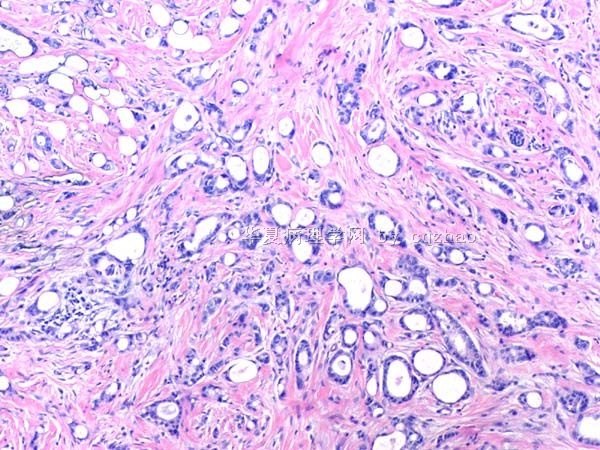
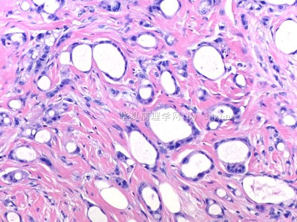
- B291244 y/f vaginal bleeding, endometrial biopsy
 图1
图1 图2
图2 图3
图3
(女,44岁,阴道不规则出血,子宫内膜活检)
40x
200x
400x
标签:乳腺癌 子宫内膜样腺癌 WT1
-
本帖最后由 于 2010-10-02 09:19:00 编辑
相关帖子
×参考诊断
腺癌。
注:描述乳腺和内膜标本的形态。根据细胞形态和免疫组化特征倾向乳腺原发。
-
本帖最后由 于 2010-10-07 21:34:00 编辑
The glands in the endometrial biopsy specimenare weakly positive for ER, focally and weakly positive for mammaglobin, and negative for GCDFP.
1. What is your thought now?
2. Is it useful for mammaglobin stain in this case?
3. What percentage of breast carcinomas is positive for GCDFP?
(内膜活检标本中,腺体呈ER弱阳性,mammaglobin局灶弱阳性,GCDFP15阴性。
1、现在考虑什么?
2、乳腺癌的GCDFP15阳性率是多少?)
mammaglobin 的敏感性和特异性也不满意,鉴别作用不大。
阳性率:
Endometrium adenocarcinoma
1/9,2/14 (positivity in the two cases was focal, weak to moderate),23/59,0/4 (using cell blocks from pleural fluid)
Breast ductal carcinoma
105/214, 9/14
References
1 Sasaki E, Tsunoda N, Hatanaka Y, et al. Breast-specific expression of MGB1/mammaglobin: an examination of 480 tumors from various organs and clinicopathological analysis of MGB1-positive breast cancers. Mod Pathol 2007; 20:208-14
2 Bhargava R, Beriwal S,Dabbs DJ Mammaglobin vs GCDFP-15: an immunohistologic validation survey for sensitivity and specificity. Am J Clin Pathol 2007; 127:103-13
3 Ciampa A, Fanger G, Khan A, et al. Mammaglobin and CRxA-01 in pleural effusion cytology: potential utility of distinguishing metastatic breast carcinomas from other cytokeratin 7-positive/cytokeratin 20-negative carcinomas. Cancer 2004; 102:368-72
4 Han JH, Kang Y, Shin HC, et al. Mammaglobin expression in lymph nodes is an important marker of metastatic breast carcinoma. Arch Pathol Lab Med 2003; 127:1330-4 FULL TEXT
5 Kataoka A, Mori M, Sadanaga N, et al. RT-PCR detection of breast cancer cells in sentinel lymph modes. Int J Oncol 2000; 16:1147-52
6 Zafrakas M, Petschke B, Donner A, et al. Expression analysis of mammaglobin A (SCGB2A2) and lipophilin B (SCGB1D2) in more than 300 human tumors and matching normal tissues reveals their co-expression in gynecologic malignancies. BMC Cancer 2006; 6:88

华夏病理/粉蓝医疗
为基层医院病理科提供全面解决方案,
努力让人人享有便捷准确可靠的病理诊断服务。
-
本帖最后由 于 2010-10-27 22:04:00 编辑
Agree above evaluation.
Both breast and current em specimens show weak ER positive.
I signed out the case:
Adenocarcinoma.
Comment: described the findings of breast and em bx specimens. Favor breast primary based on cytomophologic and IHC features.
译:
同意上述分析。
乳腺标本和现在的内膜标本均呈ER弱阳性。
我签发的报告:
腺癌。
注:描述乳腺和内膜标本的形态。根据细胞形态和免疫组化特征倾向乳腺原发。
| 以下是引用qianxun在2010-11-10 7:52:00的发言:
Dr. Zhao: I am not sure it is breast primary based on ER positive. Did you do DNA mapping for these two tumors? Thanks. Qianxun |
Thank your comment. We made the dx based on clinical hx, cytomorphology and stains. I am not 100% sure the breast primary
This is why my dx is Adenocarcinoma (see comment)
Comment: described the findings of breast and em bx specimens. Favor breast primary based on cytomophologic and IHC features
I did not do DNA mapping. Often I feel it is not very useful.
| 以下是引用cqzhao在2010-11-11 4:49:00的发言: In fact later we found this patient had other organ metastasis. It is breast ca with multiple distant metastasis. |
Journal of Gynecologic Surgery
Metastatic Breast Cancer to the Uterus and Cervix
To cite this article:
Amy C. Klostermann Eichholz, John P. Geisler, Anil K. Sood. Journal of Gynecologic Surgery. September 2003, 19(3): 145-147. doi:10.1089/104240603770191096.
Amy C. Klostermann Eichholz, John P. Geisler, Anil K. Sood. Journal of Gynecologic Surgery. September 2003, 19(3): 145-147. doi:10.1089/104240603770191096.


