| 图片: | |
|---|---|
| 名称: | |
| 描述: | |
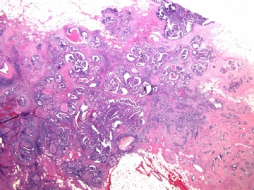
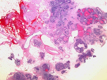
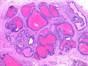
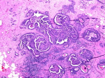
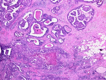
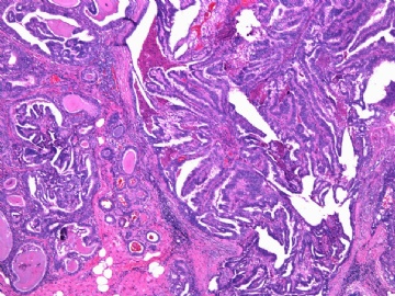
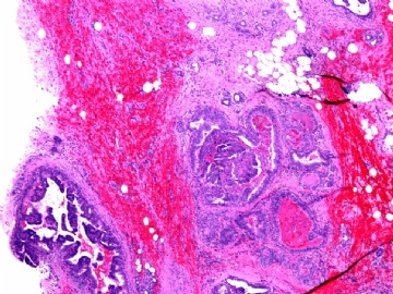
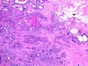
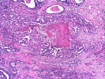
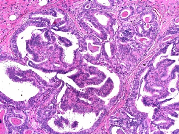
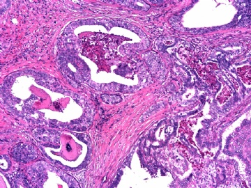
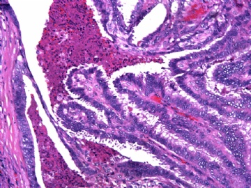
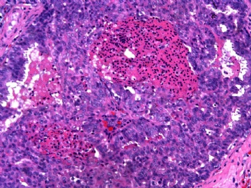
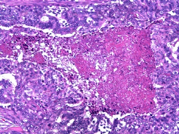
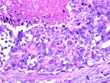
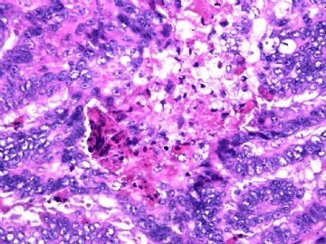
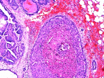
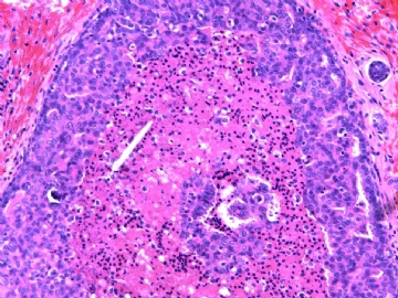
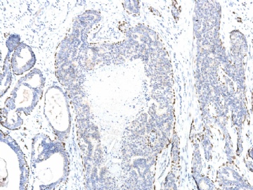
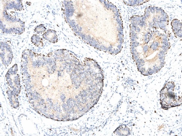
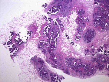
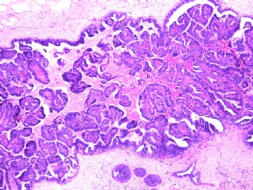
- B2906女/39岁,右乳腺肿块2.5cm,诊断?(IHC 2010-10-2)
| 姓 名: | ××× | 性别: | 年龄: | ||
| 标本名称: | 腺叶切除术 | ||||
| 简要病史: | 乳腺肿块3月,钼靶及MRI提示不规则钙化,恶性可能。 | ||||
| 肉眼检查: | 组织一块,4.5x3.5x2.5cm, 切面见不规则形肿块2.2x1.6cm, 灰白色,质地硬,有少量粉刺样物和沙砾感。 | ||||
标签:DCIS 硬化性腺病 导管内乳头状瘤 柱状细胞化生 钙化
-
本帖最后由 于 2010-10-02 12:39:00 编辑

- xljin8
相关帖子
- • 左乳癌标本乳头一个导管内的病变
- • 乳腺两个相邻导管内的病变
- • 乳腺肿物,请各位老师帮忙会诊
- • 女 46岁发现左乳腺肿块一月余
- • 求助:56岁女,左乳肿物,能排除小管癌吗?
- • 38岁乳腺(新加HE切片)
- • 乳腺肿物
- • 乳腺肿物
- • 乳腺肿块
- • 左乳肿块
×参考诊断
| 以下是引用cqzhao在2010-10-2 18:51:00的发言:
-DCIS, nuclear grade 2, solid type with comedonecrosis -DCIS is present in xx of xx submitted slides (or xx mm -microscopic measurement) -Margins are negative for DCIS; DCIS is xx mm to xx clostest margin -Intraductal papillona -sclerosing adenosis -microcalcifications associated with xx |
Dr.Zhao 建议的诊断为:
1)导管原位癌,核分级2级,实体型伴粉刺样坏死
2)导管原位癌存在于切片中的某某号(或显微镜下测量为某某mm大小);
3)组织切缘导管内癌阴性(或显微镜下测量DCIS距某方位最近切缘为 XX mm)
4)导管内乳头状瘤
5)硬化性乳腺病
6)某病变伴有微钙化
非常感谢!

- xljin8
-DCIS, nuclear grade 2, solid type with comedonecrosis
-DCIS is present in xx of xx submitted slides (or xx mm -microscopic measurement)
-Margins are negative for DCIS; DCIS is xx mm to xx clostest margin
-Intraductal papillona
-sclerosing adenosis
-microcalcifications associated with xx

