| 图片: | |
|---|---|
| 名称: | |
| 描述: | |
- 宫颈病变
-
zhongshihua 离线
- 帖子:1608
- 粉蓝豆:0
- 经验:1651
- 注册时间:2006-09-11
- 加关注 | 发消息
-
本帖最后由 于 2007-09-18 16:59:00 编辑
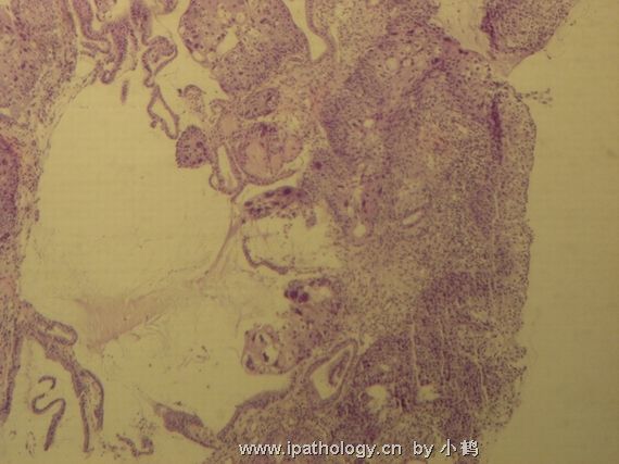
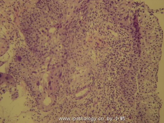
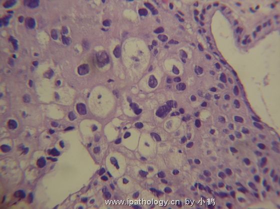
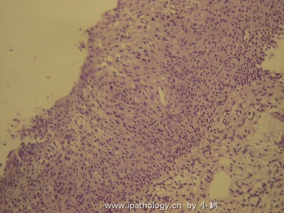
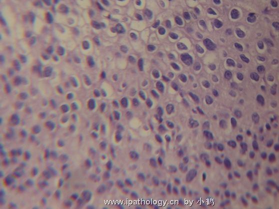
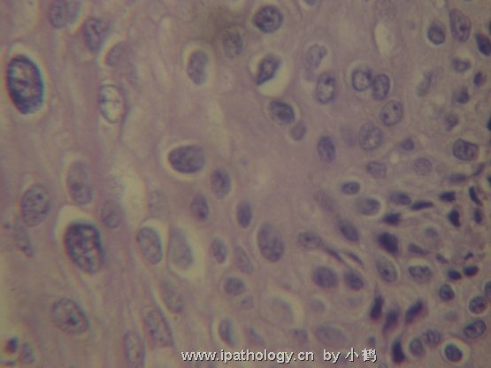
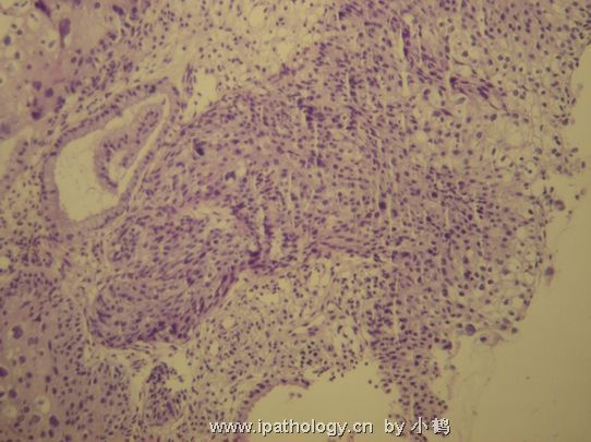
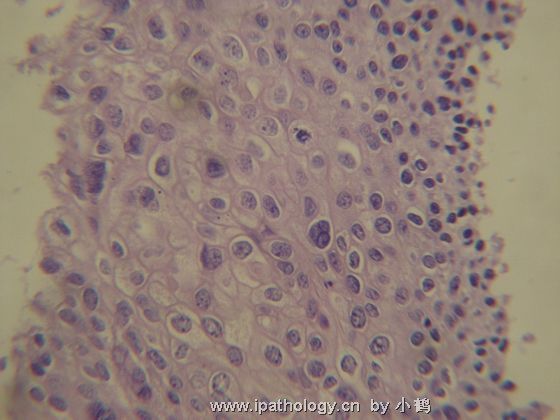
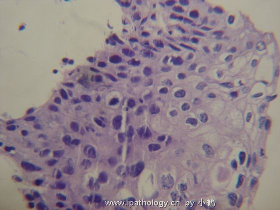
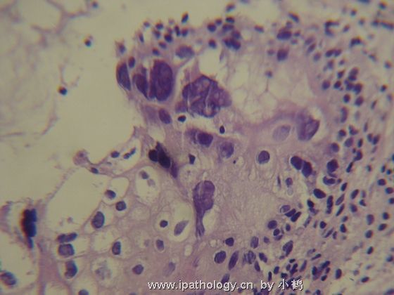
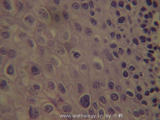
Interesting case. From the pictures postes, I did not see definitive CIN2 lesion. All I see are florid typical CIN-1 changes, which show enlarged nuclei with koliocytosis at the superficial half of the squamous epithelium. Remember, CIN-1 is superficial and intermediate cell atypia which is different from CIN2-3 which has basal cell or parabasal cell atypia. However, since CIN-1 involving endocervical glands is very rare, when you have glandular involvement, you should searching for underlying CIN2 above lesion which can co-exist with CIN-1 lesion.
So I cannot give you definite answer based on the photos provided, but provide my thought process to share for discussion.
(有趣的病例!这些图片未见明确的CIN2病变,仅见旺炽的典型的CIN-1改变--中表层核增大和控空细胞。记住二者的区别:CIN-1为表层和中层细胞不典型性,而CIN2-3为基底细胞或旁基底细胞的不典型性。然而,CIN-1极少累及宫颈腺体,如果发现累腺,应该仔细寻找有无暗藏的CIN-2,它可能与CIN-1病变共存。
因此,根据这些图片我不能明确诊断,仅提供诊断思路供讨论。abin译)

- 不坠青云之志,长怀赤子之心
-
sunxiaofeng 离线
- 帖子:98
- 粉蓝豆:5
- 经验:98
- 注册时间:2007-06-17
- 加关注 | 发消息
-
本帖最后由 于 2007-09-18 17:53:00 编辑
Dear 小鹤 ,
Thanks for your questions. As I mentioned early, CIN-1 and CIn-2-3 are two different pathologic stages with totally different morphology. If you understand HPV biology in cervix, it will be helpful in understand the morphologic changes. In CIN-1, HPV infection is in its episomal stage, in which HPV using the host protein syntheis machinary to synthesize viral protein during the squamous maturation process, including capsule protein L1 and L2 and release packaged HPV virus from the surface. That is why CIN-1 is atypia mainly in intermediate and superficial cells. You will have enlarged nuclei and koilocytosis in the top half of the epithelium. In CIN-2-3, HPV viral genome integrated into host cell genome at the basal and parabasal layer. Biologically HPV lost L1 and L2 and start to synthesize E6 and E7, two important viral oncoproteins which knock down two most important host tumor suppressor genes p53 and pRB. Therefore, morphologically CIN2-3 are basal cell atypia. Those basal cells proliferate and gradually lost maturation and replace of full epithelial layer with atypial basal-like cells. From cytology point of view, CIN-1 always have 2-3 times larger nuclei, while nuclei in CIN2-3 cells are relatively smaller than CIN-1, usually 1-2 times larger. Some of CIN2-3 cells even do not significantly increase their nuclear size. Therefore, if you have very big nuclei with para-nuclear clearance (halos) sitting in the top half of the epithelium, it is CIN-1, not CIN2-3. The most common and important differential diagnosis from CIN2-3 is not CIN-1, rather immature squamous metaplasia. In this case, immunostain with p16 is useful in differential immature squamous metaplasia (negative) from CIN2 above (positive).
{谢谢提问。如上所述,CIN-1和CIn2-3是两种不同的病理阶段,形态学完全不同。如果理解宫颈HPV的生物学,会有助于理解形态改变。
在CIN-1,HPV感染处于“附加期”(episomal stage),在鳞状上皮成熟过程中,HPV利用宿主的蛋白质合成机制来合成病毒蛋白,包括荚膜蛋白L1和L2,并且从鳞状上皮表面释放整合的HPV病毒。这可解释为什么CIN-1的异型性主要见于中表层细胞,可见鳞状上皮的上半部有增大的核和挖空细胞。
在CIN2-3,HPV病毒基因组整合到基底细胞层和旁基底细胞层的宿主细胞基因组。生物学上,HPV丢失L1和L2,开始合成E6和E7,后二者为两种重要的病毒癌蛋白,它们摧毁宿主的肿瘤抑制基因p53和pRB。因此,形态学上CIN2-3表现为基底细胞的异型性。这些基底细胞增生并逐渐失去成熟能力,并逐渐由基底样细胞取代全层上皮。
从细胞学角度,CIN-1常表现为核增大2-3倍,与CIN-1相比,CIN2-3的细胞相对较小,通常增大1-2倍。某些CIN2-3甚至没有显著的核增大。
因此,如果在表皮上半部见到非常大的核,伴核周空晕,它是CIN-1,而不是CIN2-3。
CIN2-3的最常见最重要的鉴别诊断不是CIN-1,而是不成熟鳞化。此时,p16免疫染色有助于区分不成熟鳞化(p16阴性)和CIN-2以上的病变(p16阳性)。 abin译)
问题1.有些人观点认为,只要中层出现瘤巨细胞,就要诊断为高级别病变,如果是HPV感染引起,也能诊断吗?
Wrong! it should be considered as CIN-1 when you have a very big nucleated cells with koilocytosis. (错误!如果核很大,伴挖空细胞,应为CIN-1。 abin译)
问题2.如何准确、正确的对待中空细胞的异型性?如果CIN-1伴异型明显的中空细胞,要诊断高级别吗?
No! CIN-1 cells often have para-nuclear halos which should not be taken as feature for CIN2-3. As matter of fact, CIN2-3 cells tend to lose those para-nuclear halos.(不能!CIN-1常有核周空晕,不能认为是CIN2-3的特征。实际上,CIN2-3的细胞倾向于失去核周空晕。 abin译)

- 不坠青云之志,长怀赤子之心

