| 图片: | |
|---|---|
| 名称: | |
| 描述: | |
- 宫颈病变
| 以下是引用杨斌在2007-9-18 1:18:00的发言:
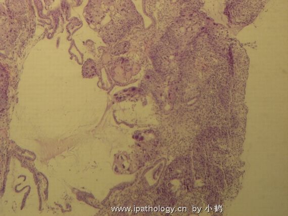
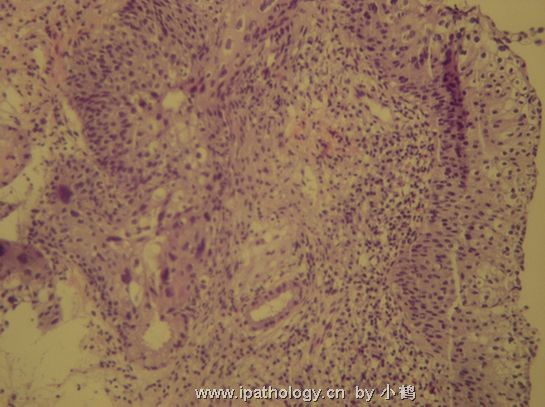
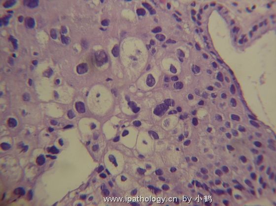
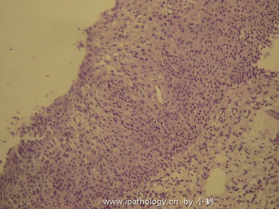
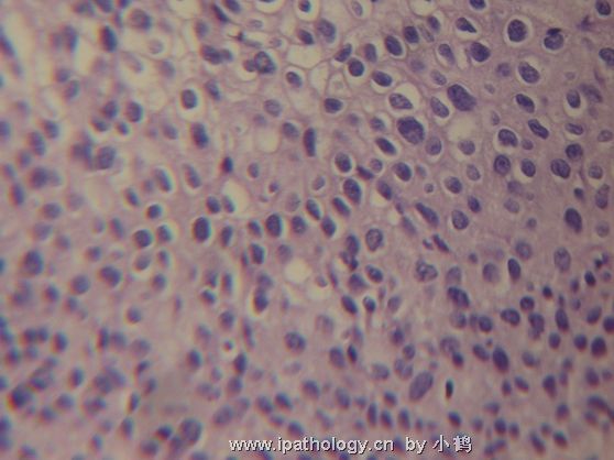
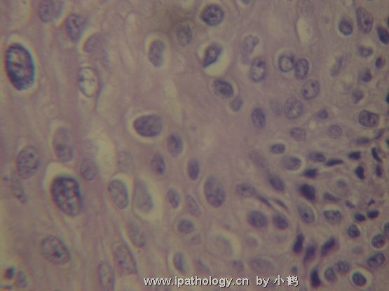
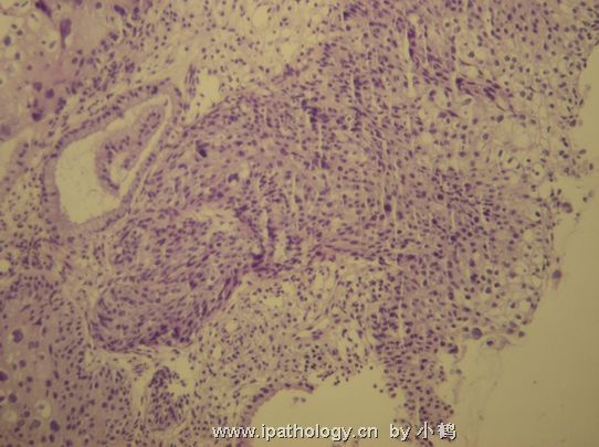
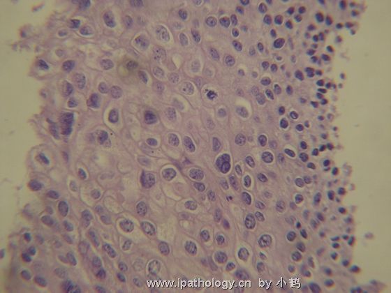
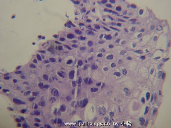
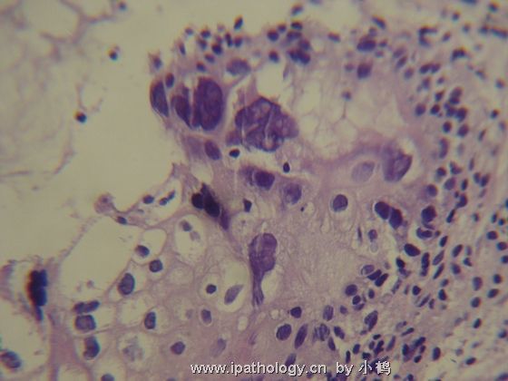
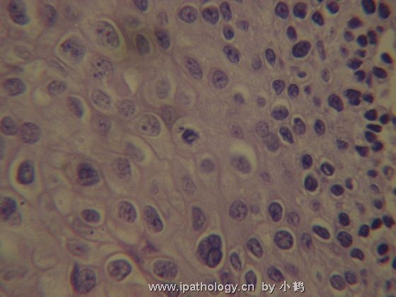
Dear 小鹤 , Thanks for your questions. As I mentioned early, CIN-1 and CIn-2-3 are two different pathologic stages with totally different morphology. If you understand HPV biology in cervix, it will be helpful in understand the morphologic changes. In CIN-1, HPV infection is in its episomal stage, in which HPV using the host protein syntheis machinary to synthesize viral protein during the squamous maturation process, including capsule protein L1 and L2 and release packaged HPV virus from the surface. That is why CIN-1 is atypia mainly in intermediate and superficial cells. You will have enlarged nuclei and koilocytosis in the top half of the epithelium. In CIN-2-3, HPV viral genome integrated into host cell genome at the basal and parabasal layer. Biologically HPV lost L1 and L2 and start to synthesize E6 and E7, two important viral oncoproteins which knock down two most important host tumor suppressor genes p53 and pRB. Therefore, morphologically CIN2-3 are basal cell atypia. Those basal cells proliferate and gradually lost maturation and replace of full epithelial layer with atypial basal-like cells. From cytology point of view, CIN-1 always have 2-3 times larger nuclei, while nuclei in CIN2-3 cells are relatively smaller than CIN-1, usually 1-2 times larger. Some of CIN2-3 cells even do not significantly increase their nuclear size. Therefore, if you have very big nuclei with para-nuclear clearance (halos) sitting in the top half of the epithelium, it is CIN-1, not CIN2-3. The most common and important differential diagnosis from CIN2-3 is not CIN-1, rather immature squamous metaplasia. In this case, immunostain with p16 is useful in differential immature squamous metaplasia (negative) from CIN2 above (positive).
Wrong! it should be considered as CIN-1 when you have a very big nucleated cells with koilocytosis. (错误!如果核很大,伴挖空细胞,应为CIN-1。 abin译)
No! CIN-1 cells often have para-nuclear halos which should not be taken as feature for CIN2-3. As matter of fact, CIN2-3 cells tend to lose those para-nuclear halos.(不能!CIN-1常有核周空晕,不能认为是CIN2-3的特征。实际上,CIN2-3的细胞倾向于失去核周空晕。 abin译) |

- If you have great talents, industry will improve them; if you have but moderate abilities, industry will supply their deficiency. 如果你很有天赋,勤勉会使其更加完美;如果你能力一般,勤勉会补足其缺陷。
| 以下是引用杨斌在2007-9-18 1:18:00的发言:
Dear 小鹤 , Thanks for your questions. As I mentioned early, CIN-1 and CIn-2-3 are two different pathologic stages with totally different morphology. If you understand HPV biology in cervix, it will be helpful in understand the morphologic changes. In CIN-1, HPV infection is in its episomal stage, in which HPV using the host protein syntheis machinary to synthesize viral protein during the squamous maturation process, including capsule protein L1 and L2 and release packaged HPV virus from the surface. That is why CIN-1 is atypia mainly in intermediate and superficial cells. You will have enlarged nuclei and koilocytosis in the top half of the epithelium. In CIN-2-3, HPV viral genome integrated into host cell genome at the basal and parabasal layer. Biologically HPV lost L1 and L2 and start to synthesize E6 and E7, two important viral oncoproteins which knock down two most important host tumor suppressor genes p53 and pRB. Therefore, morphologically CIN2-3 are basal cell atypia. Those basal cells proliferate and gradually lost maturation and replace of full epithelial layer with atypial basal-like cells. From cytology point of view, CIN-1 always have 2-3 times larger nuclei, while nuclei in CIN2-3 cells are relatively smaller than CIN-1, usually 1-2 times larger. Some of CIN2-3 cells even do not significantly increase their nuclear size. Therefore, if you have very big nuclei with para-nuclear clearance (halos) sitting in the top half of the epithelium, it is CIN-1, not CIN2-3. The most common and important differential diagnosis from CIN2-3 is not CIN-1, rather immature squamous metaplasia. In this case, immunostain with p16 is useful in differential immature squamous metaplasia (negative) from CIN2 above (positive).
非常经典的解释,学习。 |

