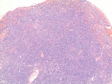
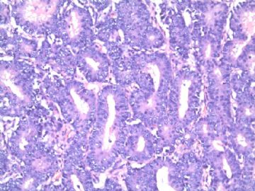
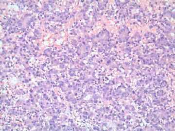
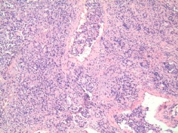
| 图片: | |
|---|---|
| 名称: | |
| 描述: | |
- 膀胱肿瘤
-
本帖最后由 于 2010-08-14 07:18:00 编辑
观察p63标记的目的有二个,1)基底细胞是否存在;2)尿路上皮分化?
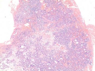
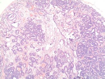
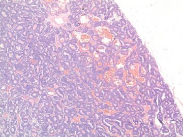
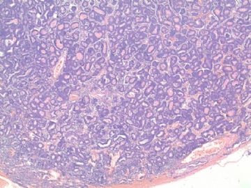
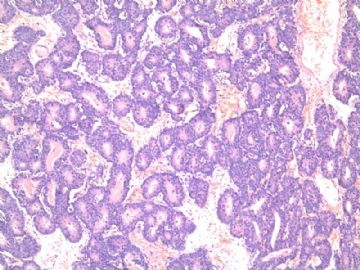
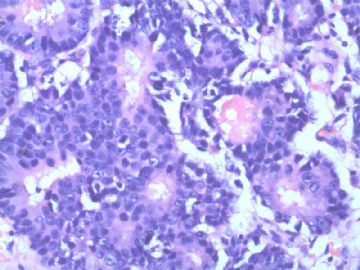
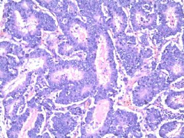
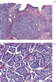
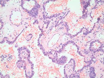
本病例主要鉴别诊断为:前列腺腺癌浸润 VS 伴有腺泡/小管分化的尿路上皮癌。
参考文献:
Huang Q, Chu PG, Lau SK, Weiss LM. Urothelial carcinoma of the urinary bladder with a component of acinar/tubular type differentiation simulating prostatic adenocarcinoma. Hum Pathol. 2004;35(6):769-73.
We report a case of an 83-year-old man with a high-grade carcinoma of the urinary bladder who underwent cystoprostatectomy. The invasive carcinoma showed mixed, morphologically distinct patterns consisting of conventional high-grade urothelial carcinoma, glandular differentiation resembling enteric type adenocarcinoma, and acinar/tubular type differentiation, morphologically similar to Gleason grade 3 prostatic adenocarcinoma.Immunohistochemical studies revealed the acinar/tubular component of the tumor to be negative for prostate-specific antigen (PSA-) and prostatic acid phosphatase(PAP-), but positive for cytokeratin 7(CK7+), cytokeratin 20(CK20+), high molecular weight cytokeratin (34 beta E12+), and thrombomodulin, consistent with origin from the bladder rather than the prostate. Although bladder carcinomas composed of mixed morphologic patterns are not uncommon, to our knowledge, the presence of acinar/tubular type features simulating prostatic adenocarcinoma in such tumors has not been described elsewhere.

- xljin8
-
本帖最后由 于 2010-08-14 07:12:00 编辑
| 34βE12 | CK7 | CK20 | CEA | PSA | PSAP | CDX2 | Villin | CA-125 | Vimentin | ||
|
Primary bladder adenocarcinoma |
+ |
+/- |
+/- |
+ |
- |
+/- |
+ |
+/- |
+/- |
- |
|
|
Prostatic adenocarcinoma |
- |
- |
+/- |
+/- |
+ |
+ |
- |
- |
- |
- |
|
|
Seminal vesicle adenocarcinoma |
|
+ |
- |
|
|
|
|
|
+ |
|
|
|
Renal cell carcinoma |
- |
- |
- |
- |
- |
- |
- |
- |
- |
+ |
|
|
Colorectal adenocarcinoma |
- |
- |
+ |
+ |
- |
- |
+ |
+ |
+/- |
- |
|
|
Endometrial adenocarcinoma |
|
+ |
- |
- |
- |
- |
- |
|
+ |
+ |
|
|
|
|
|
|
|
|
|
|
|
|
|
|

- xljin8
-
本帖最后由 于 2010-08-15 10:31:00 编辑
| 以下是引用xljin8在2010-8-12 20:29:00的发言:
观察p63标记的目的有二个,1)基底细胞是否存在;2)尿路上皮分化? 本病例主要鉴别诊断为:前列腺腺癌浸润 VS 伴有腺泡/小管分化的尿路上皮癌。 参考文献: Huang Q, Chu PG, Lau SK, Weiss LM. Urothelial carcinoma of the urinary bladder with a component of acinar/tubular type differentiation simulating prostatic adenocarcinoma类似前列腺腺癌的伴有腺泡-小管分化的膀胱肿瘤(伴腺泡/小管型分化成分类似于前列腺癌的膀胱尿路上皮癌-Dr.XLjin的翻译修改). Hum Pathol. 2004;35(6):769-73. We report a case of an 83-year-old man with a high-grade carcinoma of the urinary bladder who underwent cystoprostatectomy. The invasive carcinoma showed mixed, morphologically distinct patterns consisting of conventional high-grade urothelial carcinoma, glandular differentiation resembling enteric type adenocarcinoma, and acinar/tubular type differentiation, morphologically similar to Gleason grade 3 prostatic adenocarcinoma.我们报道了一例83岁老年病人膀胱前列腺切除术后诊断为高级别膀胱尿路癌的病例。这例浸润性癌显示混合性多种形态学亚型构成:一般的高级别尿路上皮癌;与Gleason3级前列腺癌类似的腺泡小管分化类型。(我们报道一例83岁老年男性,行膀胱前列腺切除术的高级别尿路上皮癌病例。这例浸润性癌表现为形态学上不同成分的混合,由普通高级别尿路上皮癌;类似于肠型腺癌的腺体;和形态类似于Gleason 3级前列腺癌的腺泡/小管所构成。。-Dr.XLjin的翻译修改)Immunohistochemical studies revealed the acinar/tubular component of the tumor to be negative for prostate-specific antigen (PSA-) and prostatic acid phosphatase(PAP-), but positive for cytokeratin 7(CK7+), cytokeratin 20(CK20+), high molecular weight cytokeratin (34 beta E12+),免疫组化的研究显示腺泡-小管分化的尿路上皮肿瘤,前列腺特异性抗原(PSA)和前列腺酸性磷酸酶(PAP)阴性,但是角蛋白7(CK7)、角蛋白20(CK20)、高分子量角蛋白(34bE12)阳性and thrombomodulin(血栓调节素+), consistent with origin from the bladder rather than the prostate(与前列腺相比更倾向于来源于膀胱.(免疫组化研究揭示肿瘤中的腺泡/小管成分前列腺特异性抗原(PSA)、前列腺酸性磷酸酶(PAP)均阴性,而CK7、CK20、高分子量角蛋白(34bE12)和血栓调节素阳性,与膀胱来向一致,而不符合前列腺来源。-Dr.XLjin的翻译修改) Although bladder carcinomas composed of mixed morphologic patterns are not uncommon, to our knowledge, the presence of acinar/tubular type features simulating prostatic adenocarcinoma in such tumors has not been described elsewhere.虽然由混合性形态结构组成的膀胱癌并不少见,但是在这些肿瘤中出现与前列腺腺癌类似的腺泡/小管型特征至今还未见其他文献描述。(感谢Dr.XLJin修改)
Dr.XLjin的摘要翻译: 伴腺泡/小管型分化成分类似于前列腺癌的膀胱尿路上皮癌 我们报道一例83岁老年男性,行膀胱前列腺切除术的高级别尿路上皮癌病例。这例浸润性癌表现为形态学上不同成分的混合,由普通高级别尿路上皮癌;类似于肠型腺癌的腺体;和形态类似于Gleason 3级前列腺癌的腺泡/小管所构成。免疫组化研究揭示肿瘤中的腺泡/小管成分前列腺特异性抗原(PSA)、前列腺酸性磷酸酶(PAP)均阴性,而CK7、CK20、高分子量角蛋白(34bE12)和血栓调节素阳性,与膀胱来向一致,而不符合前列腺来源。虽然由混合性形态结构组成的膀胱癌并不少见,但是在这些肿瘤中出现与前列腺腺癌类似的腺泡/小管型特征至今还未见其他文献描述。
非常感谢Dr.XLjin的修改,供我们学习参考,再次感谢。 |
-
本帖最后由 于 2010-08-15 10:47:00 编辑
学习:
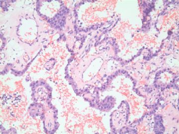
明月老师在前面的讨论已经说明:图8类似区域的图片对于诊断本例的良恶性有帮助,低级别尿路上皮肿瘤浸润区和脉管内留拴的诊断成立的前提下。
结合Dr.XLJin提供的文献和鉴别诊断资料,比较符合文献中提到的伴有混合性成分(腺泡小管分化)的低级别尿路上皮肿瘤,亦可见良性乳头状瘤样增生成分。(头脑发晕的分析,请大家原谅,作为发面教材参考 )
)
非常感谢老师们的资料和讨论!!
-
cnlzh20060 离线
- 帖子:224
- 粉蓝豆:58
- 经验:378
- 注册时间:2009-02-27
- 加关注 | 发消息
-
本帖最后由 于 2010-08-15 05:37:00 编辑
形态学:
1)浸润性低分化癌, 以筛孔和腺管为主;
2)血管内癌栓;
3)无典型的原位和浸润性泌尿上皮癌形态
4)不符合文献报道“泌尿上皮癌伴腺泡/小管分化”形态特点;
5)可见中肾样结构(见28楼);
IHC标记结果:
1)CK7-CK20- 非泌尿上皮癌免疫表型;
2)PAP-/+;AR+; P504S-/+ 倾向前列腺癌浸润/转移;
主要鉴别:膀胱原发性腺癌还是前列腺癌浸润/转移。
联系临床:前列腺癌病史?血清PSA检查?
鉴别意义:治疗策略不同;预后不同。

- xljin8
