| 图片: | |
|---|---|
| 名称: | |
| 描述: | |
- 子宫内膜
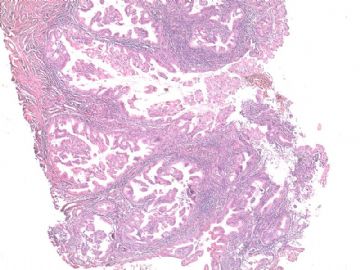
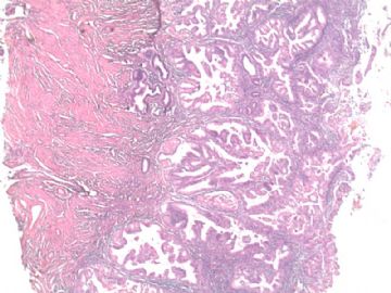
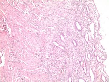
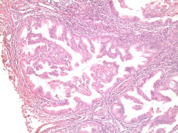
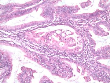
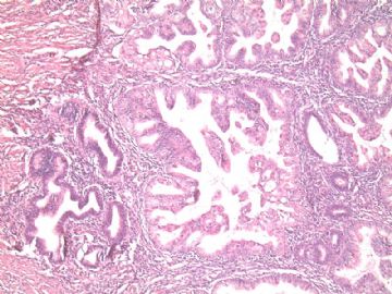
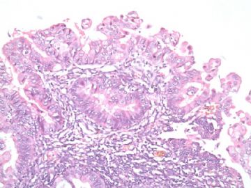
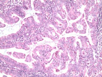
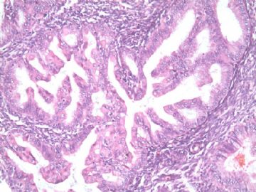
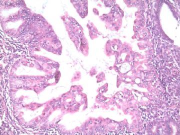
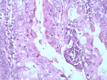
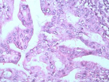
The H&E sections showc omplex papillary structure and the cells wtih mild to moderate cytologic atypia. At least it is atypical complex hyperplasia.
是不是内膜内癌就见人见智. It is true for this case. It is better to read the glass slides and know the size of focal atypical proliferation.
Based on the cytomorphology and negative P53 stain, it is not a type 2 tumor.
I favor a dx of focal endometrial adenocarcinoma, endometrioid type, FIGO grade 1, in the background of atypical complex hyperplasia.
Just for your reference.
| 以下是引用cqzhao在2010-8-20 1:03:00的发言:
The H&E sections showc omplex papillary structure and the cells wtih mild to moderate cytologic atypia. At least it is atypical complex hyperplasia. 是不是内膜内癌就见人见智. It is true for this case. It is better to read the glass slides and know the size of focal atypical proliferation. Based on the cytomorphology and negative P53 stain, it is not a type 2 tumor. I favor a dx of focal endometrial adenocarcinoma, endometrioid type, FIGO grade 1, in the background of atypical complex hyperplasia. Just for your reference. |
赵老师说:
HE切片显示复杂性乳头状结构,其细胞轻-中度异型。这至少是非典型性复杂型增生(过长)。是不是内膜内癌就见人见智? 确实如此。最好是亲阅切片,看看非典型性增生病灶的大小范围。
鉴于细胞形态和P53标记阴性,那就不是II型宫内膜肿瘤。
个人倾向诊断为局灶性(限局性)子宫内膜腺癌,宫内膜样型,FIGO I,背景病变为非典型性复杂型增生。
仅供参考。

- 王军臣
