| 图片: | |
|---|---|
| 名称: | |
| 描述: | |
- B2755关于不典型或者癌变的免疫标记的困惑,请指教(附病例)
| 姓 名: | ××× | 性别: | 女 | 年龄: | 42岁 |
| 标本名称: | 左乳肿物 | ||||
| 简要病史: | 发现左乳肿物1个月 | ||||
| 肉眼检查: | 送检组织一块,大小4×3×2cm,切面灰白灰红,局部质略硬 | ||||
-
本帖最后由 于 2010-06-28 11:32:00 编辑

- 每天前进一点点.....
相关帖子
- • 左乳肿物
- • 女性/40岁 左乳腺肿块,诊断?
- • 请教乳腺诊断
- • 导管内乳头状瘤,有癌变吗?
- • 乳腺肿物
- • 乳腺肿块,请会诊
- • 求助,乳腺肿物
- • 乳腺肿物,请大家帮忙会诊
- • 乳腺包块-请会诊
- • 左乳肿块
| 以下是引用abin在2010-7-14 23:04:00的发言:
|
-
dychocolate 离线
- 帖子:57
- 粉蓝豆:2
- 经验:65
- 注册时间:2008-07-29
- 加关注 | 发消息
| 以下是引用cqzhao在2010-6-28 15:13:00的发言:
P63 and sma stains (last two) show negative result within the areas of the ductal proliferation (no myoepithelial cells locally). It can represent UDH, ADH or DCIS. The key is to check the cytologic feature. The local proliferative cells demonstrate variable size and shape, irregular holes, more like UDH. I favor 导管内乳头状瘤with florid UDH. Suggest to stain myoepithelial markers for papillary lesions if needed. Two main point to evaluate papillary lesions: myoepithelial cells and cytology. I seldom stain 腺上皮CK5/6、CK34βE12 for papillary lesions。 |
P63和SMA染色(最后2张)显示在导管增生的区域(局部没有腺上皮)为阴性。这种情况在UDH、ADH、DCIS中都可以出现,关键是要观察细胞形态。局部增殖的细胞显示出不同的大小和形状、不规则的核,像是UDH。我更倾向于导管内乳头状瘤伴UDH。
建议如果需要的话,做肌上皮的染色来观察乳头状结构的区域,来鉴别乳头状结构的主要2点,就是肌上皮细胞和细胞学。我很少用腺上皮CK5/6、CK34βE12染色来判定乳头状结构。
| 以下是引用abin在2010-7-3 17:42:00的发言:
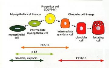
不表达这两种肌上皮标记物,并不一定没有肌上皮细胞存在。 下图有助于理解乳腺肌上皮/导管上皮的发育分化和免疫组化表达情况,还能理解为什么CK5/6有助于UDH和ADH的鉴别。 |
不表达这两种肌上皮标记物,并不一定没有肌上皮细胞存在。
This is good point.
还能理解为什么CK5/6有助于UDH和ADH的鉴别。
I never feel this stain is useful for equivocal cases (ADH vs low grade DCIS). For classic cases we do not this stain also. So ck5/6 for the differentiation is no any meaning
| 以下是引用thp12933在2010-6-29 9:13:00的发言:
谢谢各位老师。形态及排列上细胞异型性确实不大。 那为什么导管内乳头状瘤增生的细胞不表达肌上皮标记物呢?普通的导管上皮增生为什么只有腺上皮的增生而肌上皮不增生?到底存不存在ADH?
如果这样,肌上皮标记在鉴别UDH/ADH甚至DCIS中的意义何在? |
Good question.
肌上皮标记:如果乳头病变内各乳头仅有腺上皮而无第二层肌上皮,这个病变可能是乳头状癌,反之则为乳头瘤。
乳头瘤病变内局部腺上皮增生,可无肌上皮存在, 它可能是udh, adh或dcis depending the cytologic features.
It is the same as a duct with solid ductal epithelial hyperplasia. there will be no myoepithelial cells present within the ductal cells. The duct with epithelial proliferation can be udh, adh, dcis.
Hope it can help.
It is better to find a good breast path book with photos to read. It will be more easy to understand.
-
本帖最后由 于 2010-06-28 19:26:00 编辑
P63 and sma stains (last two) show negative result within the areas of the ductal proliferation (no myoepithelial cells locally). It can represent UDH, ADH or DCIS. The key is to check the cytologic feature. The local proliferative cells demonstrate variable size and shape, irregular holes, more like UDH.
I favor 导管内乳头状瘤with florid UDH.
Suggest to stain myoepithelial markers for papillary lesions if needed.
Two main point to evaluate papillary lesions: myoepithelial cells and cytology.
I seldom stain 腺上皮CK5/6、CK34βE12 for papillary lesions。


 学习了
学习了


