| 图片: | |
|---|---|
| 名称: | |
| 描述: | |
- 系统学病理及英语病理学之一
1a Chronic Pancreatitis
Chronic pancreatitis is characterized by patchy fibrous replacement of whole lobules or parts of lobules, focal fat necrosis in different stages, and chronic inflammation. Grossly, depending on the etiology and degree of injury, the gland may be slightly firm but have a normal outline, lobular pattern, and color. Severe cases may be smaller than normal, bosselated, rock-hard, and display foci of fat necrosis, calcification, or fully developed calculi.
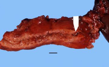
Gross: This chronically inflamed pancreas is small and scarred. The dilated pancreatic duct contains a stone (arrow). Chronic Pancreatitis with stone
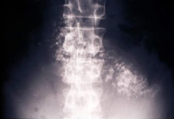
onmouseover="showMenu(this.id, 0, 1)"> X-ray: This is an example of chronic pancreatitis with marked calcification of the pancreatic parenchyma. This calcification could be demonstrated by an x-ray of the abdomen.
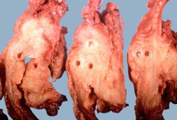
onmouseover="showMenu(this.id, 0, 1)"> Gross: These are cross sections taken through the head of the pancreas in chronic pancreatitis. The sections of pancreas which you have in your class sets were taken in a similar fashion. Notice that the dense white fibrous scarring has almost totally obliterated the lobular architecture of the pancreas.
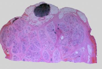
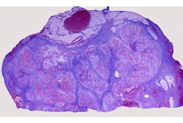
onmouseover="showMenu(this.id, 0, 1)"> This is a low magnification picture of the H&E slide of chronic pancreatitis in your class set. This particular patient had an obstructive type of pancreatitis due to carcinoma of the head of the pancreas. The lobular architecture of the pancreas is accentuated by broad bands of interstitial fibrosis.
onmouseover="showMenu(this.id, 0, 1)"> The fibrous scarring of the pancreas is highlighted with the Trichrome stain. The fibrous septa appear as blue bands surrounding the residual pancreatic lobules, which stain red.
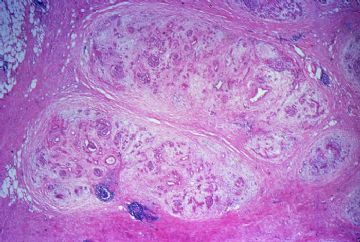
 Shows the dense interstitial fibrosis surrounding two pancreatic lobules. Note there is also an increase in intralobular fibrous tissue and there is chronic inflammation.
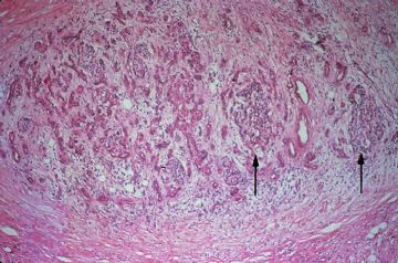
onmouseover="showMenu(this.id, 0, 1)"> The exocrine portion of the gland is most severely affected. There is almost total atrophy and fibrous replacement of pancreatic acini with relative preservation of the pancreatic ducts and islets of Langerhans (arrows). The intralobular pancreatic ductules are irregularly shaped and are embedded in chronically inflamed fibrous tissue. Several preserved islets are marked with arrows
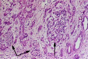
Higher magnification shows preservation of the islets of Langerhans to better advantage (arrows).

- 赚点散碎银子养家,乐呵呵的穿衣吃饭
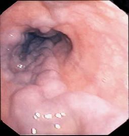
Esophageal Varices
onmouseover="showMenu(this.id, 0, 1)">

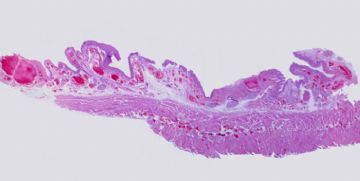
This is a photograph of the actual histologic slide in your class set. Note the markedly dilated veins in the submucosa and in the muscularis propria of the esophagus.
onmouseover="showMenu(this.id, 0, 1)">

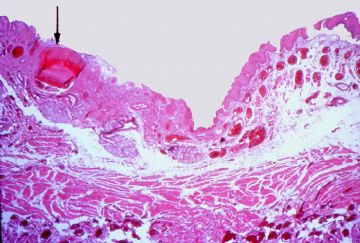
These dilated varices often cause pressure atrophy and ulceration of the overlying mucosa, as has happened in this case. The subsequent inflammation of the submucosa causes erosion of the thin wall of the varices with subsequent rupture. The large varix noted by the arrow communicates directly with the lumen and has probably ruptured in the past. It is now thrombosed.
onmouseover="showMenu(this.id, 0, 1)">

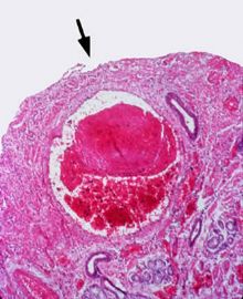
Shows thrombosis of a large submucosal varix. Note the ulceration of the overlying mucosa (arrow) and inflammation of the submucosal stroma.
onmouseover="showMenu(this.id, 0, 1)">


- 赚点散碎银子养家,乐呵呵的穿衣吃饭
Adenocarcinoma of the Pancreas
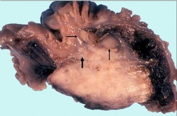
Gross: This section is taken through an adenocarcinoma of the head of the pancreas. The lesion is a firm, tan-white mass that effaces the normal lobular architecture of the pancreas. Although this carcinoma is only about 2.5 cm in diameter, it has already invaded into the wall of the duodenum (large arrow) and metastasized to regional lymph nodes (small arrows).
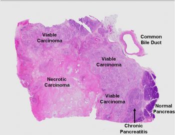
This is a low power view of the histologic slide in your class set. This is a section taken through the head of the pancreas from a patient with a ductal type of adenocarcinoma. The viable tumor is peripherally located around a central core of necrosis. Immediately adjacent to the tumor, the pancreas shows chronic pancreatitis (arrow). One normal pancreatic lobule is seen at the extreme right hand edge of the slide. The slide which you have contains a section taken through the extrapancreatic portion of the common bile duct (CD).
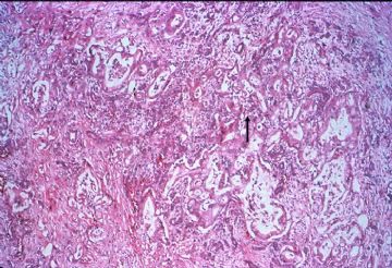
onmouseover="showMenu(this.id, 0, 1)"> This low power view shows a moderately differentiated adenocarcinoma. Irregularly shaped malignant glands are embedded in chronically inflamed fibrous tissue. Adenocarcinomas of the pancreas typically excite this type of fibrous reaction (“desmoplasia”) in the surrounding tissue. When palpated, the tumor may be extremely hard, making it virtually impossible to differentiate from chronic pancreatitis
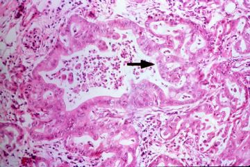
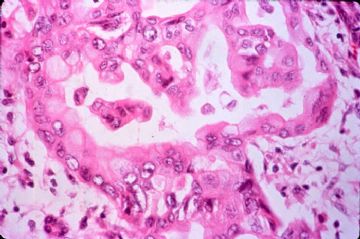
onmouseover="showMenu(this.id, 0, 1)"> This is a higher power view of a malignant gland. It is lined by pleomorphic cells which pile-up to form secondary lumina (gland-within-gland formation). Bridges of neoplastic epithelium grow across the major lumen of the gland (arrow).
 At higher magnification, note the nuclear pleomorphism of the neoplastic cells lining the glands.
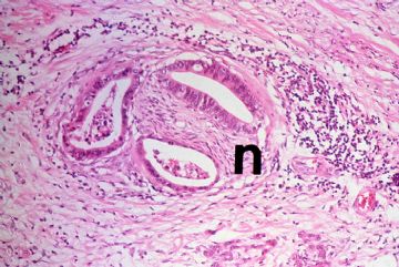
onmouseover="showMenu(this.id, 0, 1)"> Perineural invasion is extremely common in carcinoma of the pancreas. Unfortunately, infiltration into the peripancreatic fat, mesenteric vessels, duodenal wall, common bile duct, and other contiguous structures such as the stomach, spleen, portal vein, peritoneal cavity is also common. Regional lymph node metastases are almost always present at the time of diagnosis.
onmouseover="showMenu(this.id, 0, 1)">

- 赚点散碎银子养家,乐呵呵的穿衣吃饭

