| 图片: | |
|---|---|
| 名称: | |
| 描述: | |
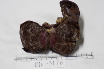
- 脾脏肿瘤?
| 姓 名: | ××× | 性别: | 男 | 年龄: | 4岁 |
| 标本名称: | 脾脏包块 | ||||
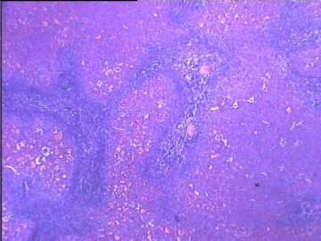
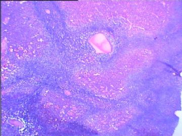
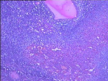
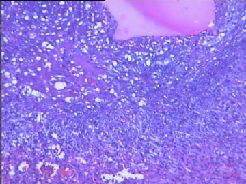
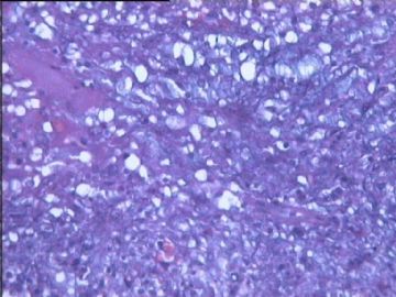
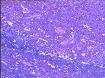
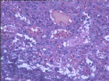
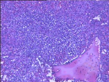
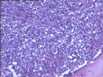
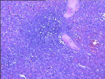
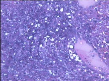
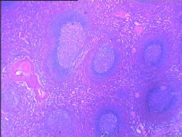
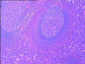
| 简要病史: | 自己无意中发现左上腹包块1周,余无其它特殊不适。CT示:脾脏下极肿块影,手术切除送检。术前周围血:白细胞、中性粒、淋巴细胞均在正常范围,Hb95g(参考值110-150g),单核细胞0.6(参考值3-8%)。(注:最后两幅图为周围正常的脾) | ||||
| 肉眼检查: | |||||
标签:
-
本帖最后由 于 2010-05-27 20:54:00 编辑
×参考诊断
-
This is a very interesting case. The pictures are not very clear. However, from the pictures we can see it is a solid tumor with irregular shaped pink vascular proliferation. There are numerous mildly dilated vascular spaces lined by bland-looking plump cells. My differential diagnosis includes littoral cell angioma, hemangioendothelialoma or others. I am favor the first. Immunohistochemical study will be very helpful. Littoral cell angioma will be positive for CD31, CD34, CD68 and lysozyme, occasionally also positive for S-100. For the hemangioendothelialoma, the only positive markers are the vascular markers, CD31, CD34 and factor 8.
Hopefully it is helpful.

