| 图片: | |
|---|---|
| 名称: | |
| 描述: | |
- 谈东风病例5 Case T0005:胃体 2cm 溃疡, 仅一张白片,该做什么?
| 姓 名: | ××× | 性别: | Male | 年龄: | 47 |
| 标本名称: | 胃镜活检 | ||||
| 简要病史: | Abdominal pain for 4 months. 7 kg weight loss. | ||||
| 肉眼检查: | Endoscopical findings: a round shallow ulcer, 2.0x1.8cm. Surrounding gastric mucosa shows atrophic changes. | ||||
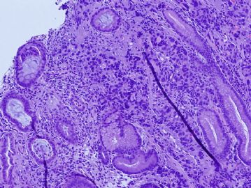
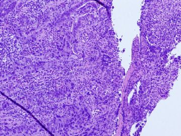
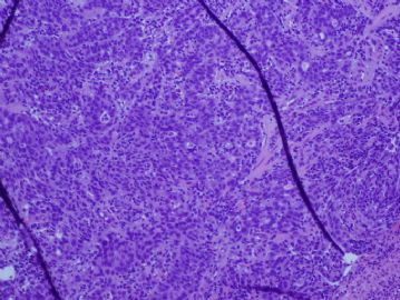
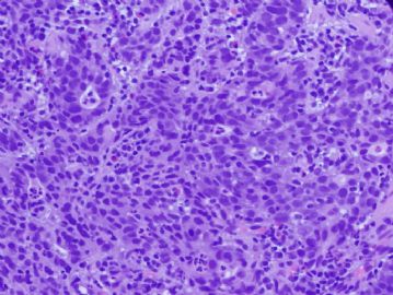
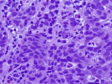
There are solid neoplastic nests in the mucosa. The tumor cells are cohensive, with scant cytoplasm, and marked intratumor lymphocytes, and background stromal lymphocytes. Morphologically, it is consistent with a poorly differentiated carcinoma, with lymphoepithelial carcinoma features.
One helpful marker to test is EBV virus. EBV associated gastric cancer (EBVAGC) was first described in early 1990 by Dr. Lawrence Weisss, who found EBV virus in lymphoepithelial carcinoma of the stomach. More and more literature have been reported, up to 16% of gastric cancer are EBV associated. In one of our recent articles, we have found EBV in other types of gastric cancer including signet ring cell carcinoma and well-differentiated intestinal type adenocarcinoma, though lymphoepithelial carcinoma type shows much higher prevalence (reference:
Characteristics of Epstein-Barr virus-associated gastric cancer: a study of 235 cases at a comprehensive cancer center in U.S.A. J Exp Clin Cancer Res. 2009 Feb 3;28:14.
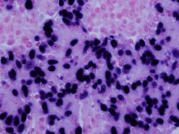
The golden standard to test EBV is in-situ hybridization (ISH), which highlights the EBV virus in the tumor nuclei. The ISH photo of the this case was shown in Figure 6. Other methods to detect EBV include PCR test and ELISA.
One of the important messages is that EBV assoicated gastric cancers seem to have a better clinical outcome compared to non-EBV associated gastric cancer.
-
本帖最后由 于 2010-05-21 03:48:00 编辑
| 以下是引用谈东风在2010-5-11 0:22:00的发言:
Hint:
Sheets of solid, cohensive neoplastic cells in a background of chronic atrophic gastritis (see intestinal metaplasia in the background). Another important feature: remarkable intratumor lymphocytes!! |
翻译:谈老师的提示
在慢性萎缩性胃炎(看背景上的肠化)的背景上见到成片实性的、粘附性的肿瘤细胞
另一个重要特点:明显的肿瘤间的淋巴细胞
-
aishanshan 离线
- 帖子:6
- 粉蓝豆:1
- 经验:6
- 注册时间:2010-05-19
- 加关注 | 发消息
There are solid neoplastic nests in the mucosa. The tumor cells are cohensive, with scant cytoplasm, and marked intratumor lymphocytes, and background stromal lymphocytes. Morphologically, it is consistent with a poorly differentiated carcinoma, with lymphoepithelial carcinoma features.
在粘膜内可见实性的肿瘤细胞巢。肿瘤细胞具有粘附性,胞浆稀少,伴有明显的肿瘤间淋巴细胞及间质淋巴细胞背景。形态学符合低分化癌,伴有淋巴上皮样癌特征。
One helpful marker to test is EBV virus. EBV associated gastric cancer (EBVAGC) was first described in early 1990 by Dr. Lawrence Weisss, who found EBV virus in lymphoepithelial carcinoma of the stomach. More and more literature have been reported, up to 16% of gastric cancer are EBV associated. In one of our recent articles, we have found EBV in other types of gastric cancer including signet ring cell carcinoma and well-differentiated intestinal type adenocarcinoma, though lymphoepithelial carcinoma type shows much higher prevalence (reference:
Characteristics of Epstein-Barr virus-associated gastric cancer: a study of 235 cases at a comprehensive cancer center in U.S.A. J Exp Clin Cancer Res. 2009 Feb 3;28:14.
EBV病毒的检测是一个有用的标记物。EBV相关胃癌(EBVAGC)首先由Lawrence Weisss于上世纪九十年代早期首先报道。他在胃的淋巴上皮样癌中发现了EBV病毒。越来越多的文献报道,胃癌中EBV相关胃癌的比例达到16%。在我们最近的一篇文章中,我们在其他类型的胃癌中也发现了了EBV病毒,包括印戒细胞癌和高分化肠型腺癌,当然淋巴上皮样癌显示出了更高的比例。
The golden standard to test EBV is in-situ hybridization (ISH), which highlights the EBV virus in the tumor nuclei. The ISH photo of the this case was shown in Figure 6. Other methods to detect EBV include PCR test and ELISA.
检测EBV的金标准是原位杂交(ISH),它可以显示细胞核中的EBV病毒。Figure 6显示了本例的原位杂交照片。检测EBV病毒的其他方式有PCR和ELISA。
One of the important messages is that EBV assoicated gastric cancers seem to have a better clinical outcome compared to non-EBV associated gastric cancer.
EBV相关胃癌的预后比非EBV相关胃癌好。
-
本帖最后由 于 2010-05-24 12:02:00 编辑
| 以下是引用knight在2010-5-22 0:23:00的发言: There are solid neoplastic nests in the mucosa. The tumor cells are cohensive, with scant cytoplasm, and marked intratumor lymphocytes, and background stromal lymphocytes. Morphologically, it is consistent with a poorly differentiated carcinoma, with lymphoepithelial carcinoma features. 在粘膜内可见实性的肿瘤细胞巢。肿瘤细胞具有粘附性,胞浆稀少,伴有明显的肿瘤间淋巴细胞及间质淋巴细胞背景。形态学符合低分化癌,伴有淋巴上皮样癌特征。 One helpful marker to test is EBV virus. EBV associated gastric cancer (EBVAGC) was first described in early 1990 by Dr. Lawrence Weisss, who found EBV virus in lymphoepithelial carcinoma of the stomach. More and more literature have been reported, up to 16% of gastric cancer are EBV associated. In one of our recent articles, we have found EBV in other types of gastric cancer including signet ring cell carcinoma and well-differentiated intestinal type adenocarcinoma, though lymphoepithelial carcinoma type shows much higher prevalence (reference: Characteristics of Epstein-Barr virus-associated gastric cancer: a study of 235 cases at a comprehensive cancer center in U.S.A. J Exp Clin Cancer Res. 2009 Feb 3;28:14.
The golden standard to test EBV is in-situ hybridization (ISH), which highlights the EBV virus in the tumor nuclei. The ISH photo of the this case was shown in Figure 6. Other methods to detect EBV include PCR test and ELISA.
One of the important messages is that EBV assoicated gastric cancers seem to have a better clinical outcome compared to non-EBV associated gastric cancer. 感谢Dr.Knight! |

