| 图片: | |
|---|---|
| 名称: | |
| 描述: | |
- 谈东风病例5 Case T0005:胃体 2cm 溃疡, 仅一张白片,该做什么?
| 姓 名: | ××× | 性别: | Male | 年龄: | 47 |
| 标本名称: | 胃镜活检 | ||||
| 简要病史: | Abdominal pain for 4 months. 7 kg weight loss. | ||||
| 肉眼检查: | Endoscopical findings: a round shallow ulcer, 2.0x1.8cm. Surrounding gastric mucosa shows atrophic changes. | ||||
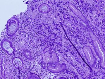
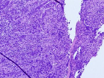
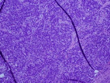
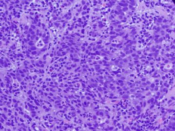
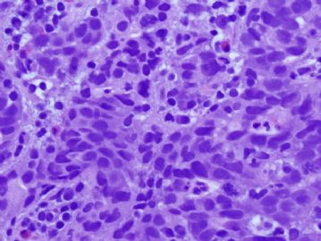
There are solid neoplastic nests in the mucosa. The tumor cells are cohensive, with scant cytoplasm, and marked intratumor lymphocytes, and background stromal lymphocytes. Morphologically, it is consistent with a poorly differentiated carcinoma, with lymphoepithelial carcinoma features.
One helpful marker to test is EBV virus. EBV associated gastric cancer (EBVAGC) was first described in early 1990 by Dr. Lawrence Weisss, who found EBV virus in lymphoepithelial carcinoma of the stomach. More and more literature have been reported, up to 16% of gastric cancer are EBV associated. In one of our recent articles, we have found EBV in other types of gastric cancer including signet ring cell carcinoma and well-differentiated intestinal type adenocarcinoma, though lymphoepithelial carcinoma type shows much higher prevalence (reference:
Characteristics of Epstein-Barr virus-associated gastric cancer: a study of 235 cases at a comprehensive cancer center in U.S.A. J Exp Clin Cancer Res. 2009 Feb 3;28:14.
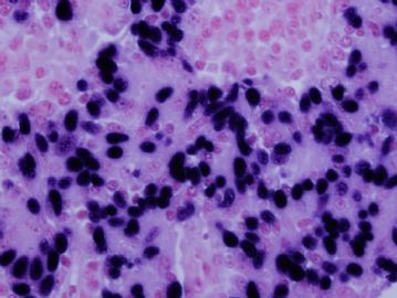
The golden standard to test EBV is in-situ hybridization (ISH), which highlights the EBV virus in the tumor nuclei. The ISH photo of the this case was shown in Figure 6. Other methods to detect EBV include PCR test and ELISA.
One of the important messages is that EBV assoicated gastric cancers seem to have a better clinical outcome compared to non-EBV associated gastric cancer.
-
本帖最后由 于 2010-05-21 03:48:00 编辑
-
yanjinputao 离线
- 帖子:49
- 粉蓝豆:408
- 经验:51
- 注册时间:2010-11-09
- 加关注 | 发消息
-
watcher035 离线
- 帖子:482
- 粉蓝豆:8
- 经验:767
- 注册时间:2010-08-14
- 加关注 | 发消息
-
wangjh1985 离线
- 帖子:2
- 粉蓝豆:1
- 经验:2
- 注册时间:2009-11-17
- 加关注 | 发消息

