| 图片: | |
|---|---|
| 名称: | |
| 描述: | |
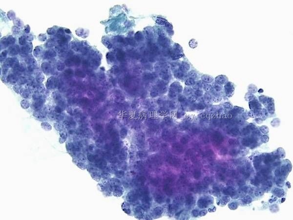
- case1 宫颈小细胞癌; case2宫颈ADC; case 3 50 y/f月经增多
I showed a case here a few days ago. It was a very complicated law-suit case and I cannot find more photos now. So I deleted it because it is not good for education.Sorry for that.(几天前我贴了一个病例在这里。那是一个非常复杂的法律诉讼病例,由于我没有更多的图片,对于教学不是很好所以我删除了它。为此我深感抱歉。)
Our fellow showed an interesting case. I put here for your review.(我们的住院医有一个很有趣的病案,我贴在这里一起分享)
42 y women with LMP 5 days ago and no previous Pap history (女性,42岁,末次月经5天前,既往无巴氏检查)
-
本帖最后由 于 2010-05-07 07:28:00 编辑
-
kangwang2010 离线
- 帖子:389
- 粉蓝豆:13
- 经验:590
- 注册时间:2010-03-04
- 加关注 | 发消息
| 以下是引用cqzhao在2010-3-13 16:33:00的发言:
When we read Pap, we should consider the patients, clinical situation, clinical managment, histology et al. These also can make the Pap more interesting. Some one can make a list for the differential diagnosis of small cell carcinoma in Pap test. Thanks, cz |
-
本帖最后由 于 2010-03-19 16:28:00 编辑
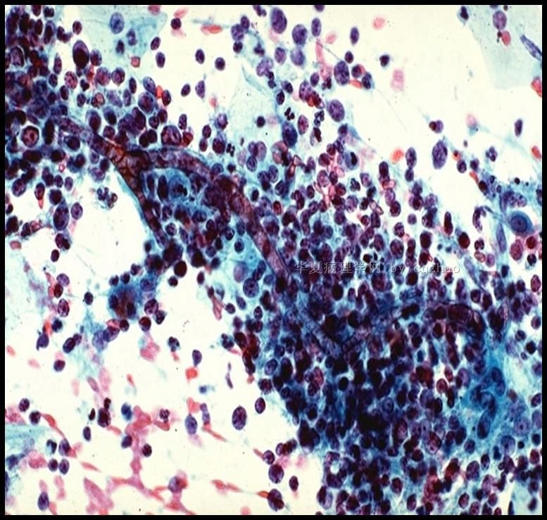
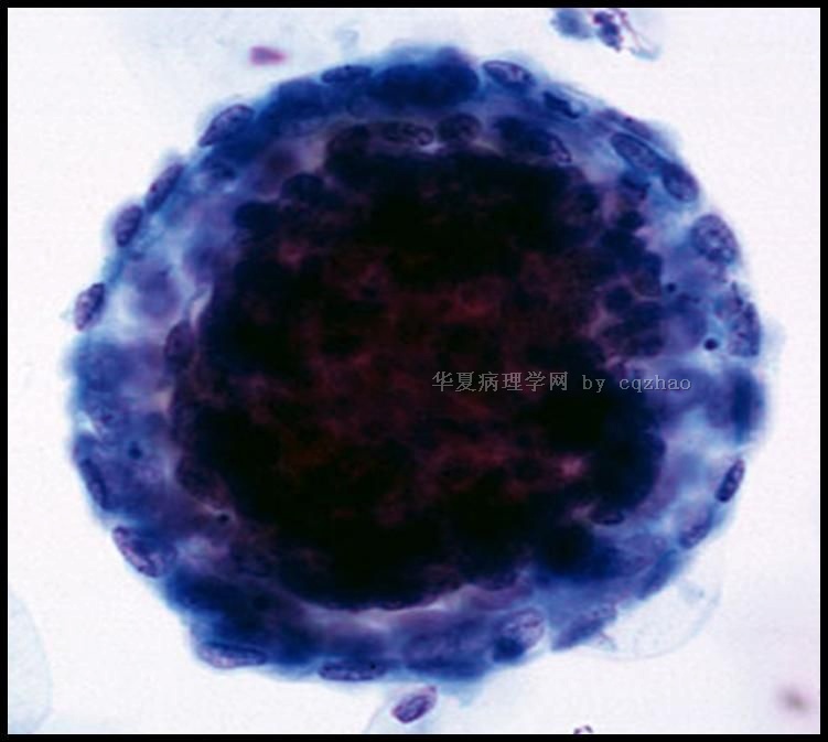
Follicular cervicitis
Lymphocytic cervicitis (lymphoid follicles in the subepithelial areas)
Cytology: mature and reactive lymphoid cells, macrophages w tingible bodies
~50% of the cases of follicular cervicitis associated with Chlamydia; also atrophy
Sometimes difficult to recognize in LBGS
– Small lymphoid cells in aggregates or dispersed
– Capillaries from the germinal center
– Should be distinguished for malignant lymphomas, histocytes, endometrial cells, and metastatic tumor cells
滤泡性宫颈炎
淋巴细胞性宫颈炎(上皮下存在淋巴滤泡)
细胞学:可见成熟的反应性淋巴细胞及巨噬细胞,约有50%衣原体相关的滤泡性炎可见可染小体(巨噬细胞吞噬淋巴细胞形成);
——聚集成堆或分散排列的小淋巴样细胞;
——有时可见生发中心来源的微血管;
——应该和恶性淋巴瘤、组织细胞、子宫内膜细胞和转移性肿瘤细胞相区分
名称:图1
描述:图1
-
本帖最后由 于 2010-03-19 16:13:00 编辑
Benign endometrial cells
Condensed stromal cells surrounded by paler epithelial cells ("exodus balls")
Closely packed epithelial cells w small hyperchromatic bean-shaped nuclei
Nuclear molding,variation in shape and size, apoptotic bodies
Cytoplasm scant, small vacuoles w engulfed PMN
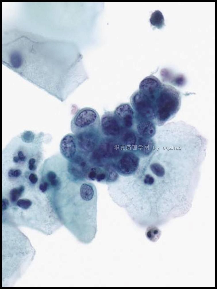
1. Endometrial ball (exodus, present in LMP 6-10 days)
2. em cells
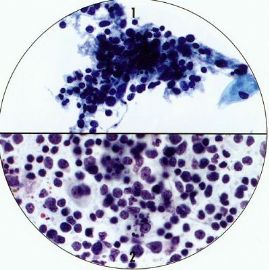
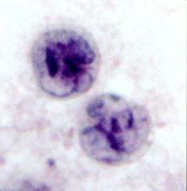
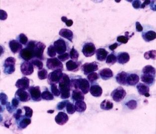
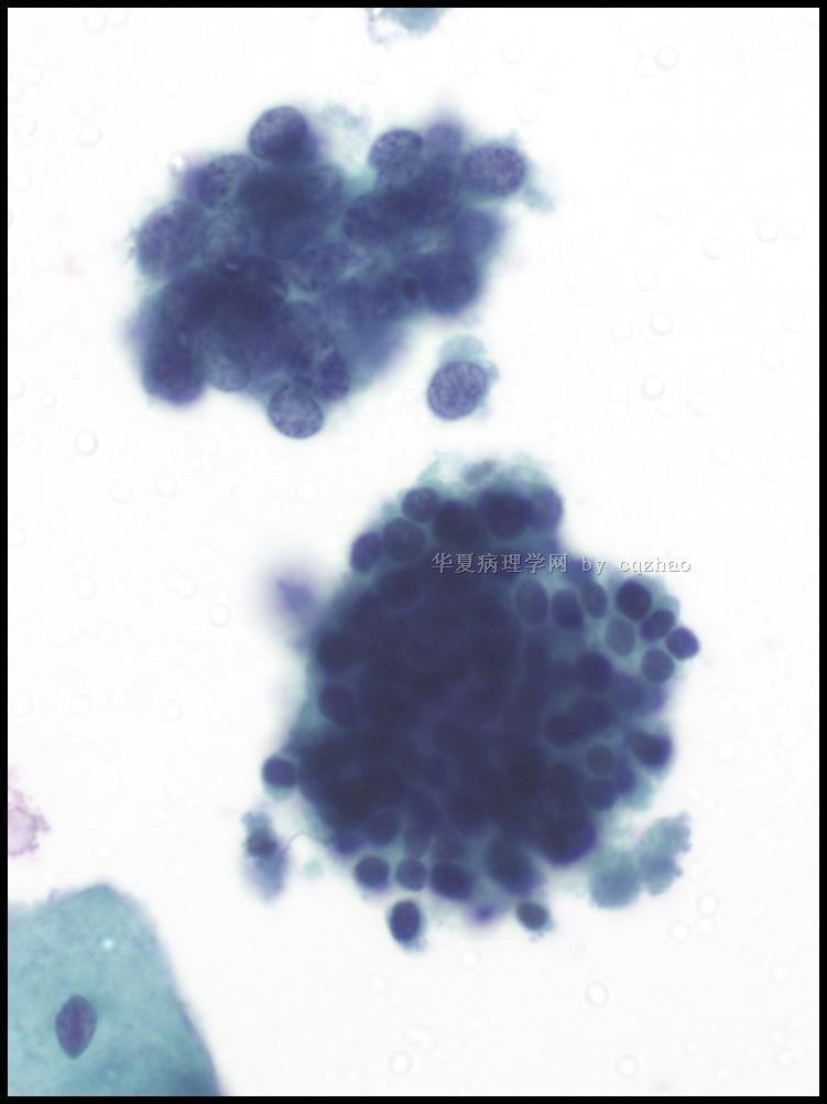
3. The case above: two clusters: upper small cell ca; low normal em cell
良性子宫内膜细胞
浓集的间质细胞周围围以栅状排列的上皮细胞(基质球)
密集成堆的上皮细胞,细胞小而深染,豆瓣形胞核,核扭曲,大小和形状多变,有时可见凋亡小体;胞质稀少,多形核细胞内可见卷入的小空泡
1、 子宫内膜基质球(LMP 6-10子宫内膜剥脱)
2、 子宫内膜细胞
3、 以上讨论本病例:两簇细胞,上面为小细胞癌,下面为正常子宫内膜细胞。
名称:图1
描述:图1
名称:图2
描述:图2
名称:图3
描述:图3
-
本帖最后由 于 2010-03-19 16:14:00 编辑
HSIL
Sheets, syncytia, single cells of variable size
Variable nuclear size, hyperchromasia, irregular membranes
Nucleoli absent (except glandular involvement)
Immature lacy or dense cytoplasm
HSIL
大小多变的片状、合胞体样或单个散在排列的细胞
胞核大小也存在较大变数,胞核深染,核膜不规则
无核仁(除外累及腺体病例)
不成熟的花边状或稠密胞质
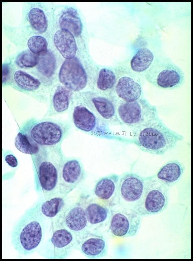
名称:图1
描述:图1
名称:图2
描述:图2
-
本帖最后由 于 2010-03-19 16:14:00 编辑
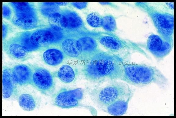
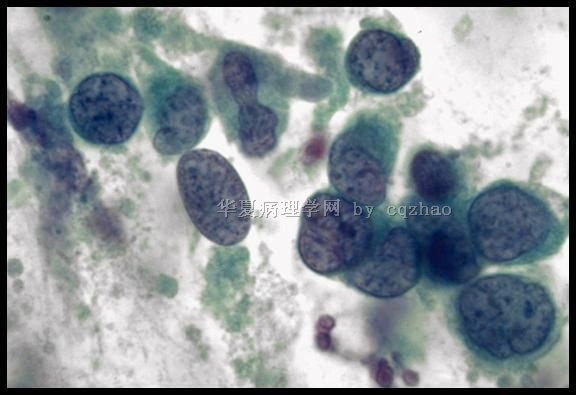
Small Cell Squamous Cell Ca
Well-defined nests of basaloid-type cells resembling small cell neuroendocrine carcinoma, but with more cytoplasm, coarser chromatin and prominent nucleoli; 60% also have SIL
Can show mixed pattern with small and non-small cell differentiation
p63 +; neuroendocrine markers -
f1 : non-keratinizing squamous cell carcinoma
f2: poorly differentiated squamous cell carcinoma
小细胞鳞癌
分化好的基底细胞样型鳞癌和小细胞神经内分泌癌很相像,但前者胞质较丰富、染色质粗糙、核仁明显,并且60%的病例可见SIL细胞
可表现为小细胞和非小细胞混合型,p63 +,神经内分泌标记-
1图:非角化性鳞癌
2图:低分化鳞癌
名称:图1
描述:图1
名称:图2
描述:图2
-
本帖最后由 于 2010-03-19 16:15:00 编辑
Small Cell Carcinoma of the Uterine Cervix
Cases containing RBCs and clusters of small hyperchromatic cells should not be screened at low power assuming they are exfoliated endometrial cells
Small cell carcinoma is HPV18+ so hrHPV co-testing can help (+in endocervical, -in endometrial)
宫颈小细胞癌
含有红细胞大小的深染小簇状的病例,低倍镜下不要认为是剥脱的子宫内膜而漏诊
小细胞癌HPV18+,所以hrHPV和细胞学共同筛查有帮助作用(+提示子宫颈来源问题,—子宫内膜)
-
本帖最后由 于 2010-03-19 16:16:00 编辑
Finally I complete this Pap case presentation and discussion here.
I hope whoever reviewed this case think about what you learn from this case. You may say I have learned how to make interpretation of cervical samll cell carcinoma in Pap. I do not think so. You may never meet one case of small cell ca of cervix in Pap for all of your life practice even though you may be young. The purpose I present the case here is to let more young pathologists know to make interpretation of the Pap or even surgical pathology. We need to think all clinical information and have open mind for all possible differential diagnosis. You can be excellent to read Pap smear, but that do not mean you are a good pathologist or cytopathologist. Cytotechnologists may have better skill to screen Pap smears, but their knowledge and 思维方式limite them as pathologists, generally speaking. I have no any meaning here to 冒犯 our Chenise cytotechnicians if we have some technicians here.
Any way I feel deserved if some ones can learn to use logic and 严谨的思维方式来分析和解读你的病例。
Thank all people who read or attended discussion for this case.
Thank God I am done for this case.
最后我完成了本例巴氏涂片病例的介绍和讨论。
我希望看过此病例的同仁都能回想一下你从中学到了什么。也许你会说:我学会了在巴氏涂片中判读宫颈小细胞癌。我不这样认为。即使你们依然年轻,但也许你从业一生也遇不到一例宫颈小细胞癌。我展示此病例的目的是想让更多的病理医生了解如何去判读巴氏涂片或如何去做外科病理学。我们必须搜集所有临床信息、开动脑筋想到所有可能的鉴别诊断,也许你看巴氏涂片很厉害,但并不意味着你一定是位优秀的病理医生或细胞病理医生。通常来讲,细胞技术员筛查巴氏涂片技术可以不断提高,但他们的知识范围和思维方式限制了他们成为病理医生的可能。这里我并无意冒犯我们中国的细胞技术员,如果这里有技师的话。
我们应该学会以逻辑的、严谨的思维方式来分析和解读病例,这方面无论怎样做都不为过。
谢谢所有浏览或参与讨论的同仁。
感谢上帝,我终于完成了这一病例的所有讨论。(感谢上帝,我终于翻译完了。青青子衿)
宫颈小细胞癌的鉴别诊断很多,几乎所有可以发生于宫颈并且含有小蓝细胞成分的病变都应该与之相互区分。除了以上赵老师分别列出的以外,还有肉瘤(尤其是子宫内膜间质肉瘤)、恶黑、白血病等等。另外,我们诊断原发性宫颈小细胞癌时一定要除外转移癌,尤其是肺源性小细胞癌,结合临床病史非常重要。
以淋巴瘤为主的淋巴造血系统病变
最应该和淋巴瘤或白血病相鉴别的应该是滤泡性宫颈炎,不仅因为二者有很多相似之处,更因为二者性质完全不同,这是原则性问题。关于这二者的鉴别诊断,我们前面有过讨论。参见以下:
1http://www.ipathology.cn/forum/forum_display.asp?classcode=108&keyno=193330&pageno=2
宫颈淋巴瘤常是全身性或系统播散性病变的一部分。记得有位老前辈曾经和我们说过:淋巴瘤的特点就是细胞分布象撒出去的一把黄豆,也就是弥漫、散布、失粘附,排列方式是其最大特点。细胞学特点:单个散在细胞群或单型群落,细胞大小相对一致,显著核仁、不规则核轮廓及粗颗粒状染色质。
图1:滤泡性宫颈炎;图2:淋巴瘤;图3:小细胞癌


