| 图片: | |
|---|---|
| 名称: | |
| 描述: | |
- 头痛脑脊液检查
Our fellow showed a case last week. I think it is interesting. Send here as a chinese new year gift for some ones interested.
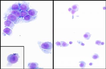
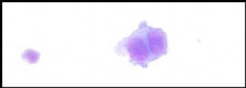
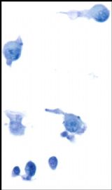
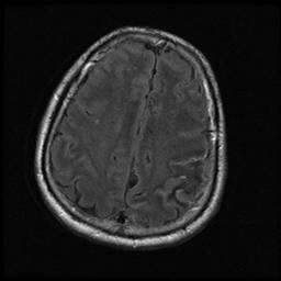
about 50 y women with headach, CSF cytology exam. paitent has no previous malignant history.
-
本帖最后由 于 2010-02-13 10:36:00 编辑
-
本帖最后由 于 2010-03-07 23:04:00 编辑
看到再次送检的CSF涂片,仍然考虑为恶性。
原发、继发?
癌、非癌?
图中可见到个别细胞核明显偏位、胞质中见到不光滑的空泡,核分裂活跃,细胞核大小不一,核仁明显,似乎还能见到少许细胞邻接和排列趋势。
考虑为转移性腺癌,建议:查消化道;尚不全排除原发可能,如果永远也发现不了原发灶的话。
但是,有没有可能是造血系统疾病呢?很多细胞的那张图(含两个分裂像的)猛一看象是白血病。会不会是腔性弥漫大B细胞淋巴瘤?纯属猜测,能用流式细胞仪多做几项标记看看就好了。

- “人生没有彩排,每一天都是现场直播”
Quickly review above interpretation, suggestion, common.
We are pathologists. It is good for us to make some suggestion. However it is important for us to provide more information or more specific diagnosis for clinic.
For example we make the diagnosis, suspicous malignancy, ? metastatic adenocarcinoma et al. What do yo want to clinicians to do based on our cytology diagnosis.
Can we do some stains for this case?


